Bishistidyl Heme Hexacoordination, a Key Structural Property in Drosophila Melanogaster Hemoglobin
De Sanctis, D., Dewilde, S., Vonrhein, C., Pesce, A., Moens, L., Ascenzi, P., Hankeln, T., Burmester, T., Ponassi, M., Nardini, M., Bolognesi, M.(2005) J Biol Chem 280: 27222
- PubMed: 15917230
- DOI: https://doi.org/10.1074/jbc.M503814200
- Primary Citation of Related Structures:
2BK9 - PubMed Abstract:
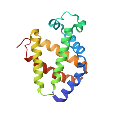
Hemoglobins at high concentration have been isolated long ago from some insect larvae living in hypoxic environments. Conversely, a monomeric hemoglobin has been discovered recently in the fruit fly Drosophila melanogaster as intracellular protein expressed both in larvae and in the adult fly. Such a finding indicates that the oxygen supply in insects may be more complex than previously thought, relying not only on O2 diffusion through the tubular tracheal system, but also on carrier-mediated transport and storage. We present here the crystal structure of recombinant D. melanogaster hemoglobin at 1.20 A resolution. Spectroscopic data show that the protein displays a hexacoordinated heme, whose axial ligands are the proximal and distal His residues. Such bis-His ligation of the heme has sizable effects on the protein local structure. Three protein matrix cavities, comparable in size but not in topological locations with those of sperm whale myoglobin, are spread through the protein matrix; one of these can host a xenon atom. Additionally, D. melanogaster hemoglobin binds one molecule of 3-(cyclohexylamino)propanesulfonic acid (CAPS) buffer at a surface pocket, next to the EF hinge. Despite the high resolution achieved, no sequence/structure features specifically supporting the heme hexa- to pentacoordination transition required for diatomic ligand binding could be recognized.
Organizational Affiliation:
Department of Physics, National Institute for the Physics of Matter (NFM), University of Genova, Genova I-16146, Italy.