2B61

Crystal Structure of Homoserine Transacetylase


Experimental Data Snapshot

  • Method: X-RAY DIFFRACTION
  • Resolution: 1.65 Å
  • R-Value Free: 0.187 
  • R-Value Work: 0.163 
  • R-Value Observed: 0.164 

wwPDB Validation   3D Report Full Report


This is version 1.3 of the entry. See complete history


Literature

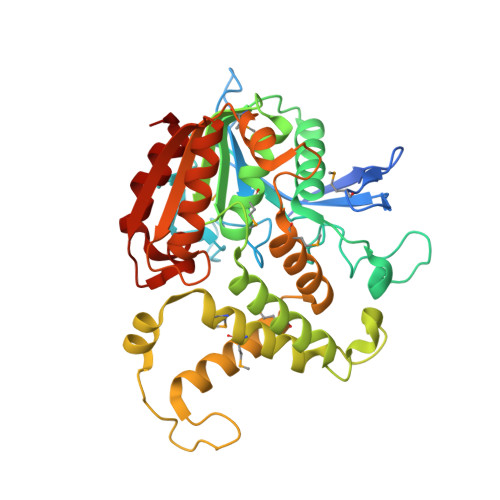
Crystal Structure of Homoserine Transacetylase from Haemophilus influenzae Reveals a New Family of alpha/beta-Hydrolases

Mirza, I.A.Nazi, I.Korczynska, M.Wright, G.D.Berghuis, A.M.

(2005) Biochemistry 44: 15768-15773

  • DOI: https://doi.org/10.1021/bi051951y
  • Primary Citation of Related Structures:  
    2B61

  • PubMed Abstract: 

    Homoserine transacetylase catalyzes one of the required steps in the biosynthesis of methionine in fungi and several bacteria. We have determined the crystal structure of homoserine transacetylase from Haemophilus influenzae to a resolution of 1.65 A. The structure identifies this enzyme to be a member of the alpha/beta-hydrolase structural superfamily. The active site of the enzyme is located near the end of a deep tunnel formed by the juxtaposition of two domains and incorporates a catalytic triad involving Ser143, His337, and Asp304. A structural basis is given for the observed double displacement kinetic mechanism of homoserine transacetylase. Furthermore, the properties of the tunnel provide a rationale for how homoserine transacetylase catalyzes a transferase reaction vs hydrolysis, despite extensive similarity in active site architecture to hydrolytic enzymes.


  • Organizational Affiliation

    Department of Biochemistry, McGill University, Montreal, Quebec, Canada H3A 1A4.


Macromolecules
Find similar proteins by:  (by identity cutoff)  |  3D Structure
Entity ID: 1
MoleculeChains Sequence LengthOrganismDetailsImage
Homoserine O-acetyltransferase377Haemophilus influenzaeMutation(s): 8 
Gene Names: metXmet2
EC: 2.3.1.31
UniProt
Find proteins for P45131 (Haemophilus influenzae (strain ATCC 51907 / DSM 11121 / KW20 / Rd))
Explore P45131 
Go to UniProtKB:  P45131
Entity Groups  
Sequence Clusters30% Identity50% Identity70% Identity90% Identity95% Identity100% Identity
UniProt GroupP45131
Sequence Annotations
Expand
  • Reference Sequence
Small Molecules
Modified Residues  1 Unique
IDChains TypeFormula2D DiagramParent
MSE
Query on MSE
A
L-PEPTIDE LINKINGC5 H11 N O2 SeMET
Experimental Data & Validation

Experimental Data

  • Method: X-RAY DIFFRACTION
  • Resolution: 1.65 Å
  • R-Value Free: 0.187 
  • R-Value Work: 0.163 
  • R-Value Observed: 0.164 
  • Space Group: P 31 2 1
Unit Cell:
Length ( Å )Angle ( ˚ )
a = 85.264α = 90
b = 85.264β = 90
c = 120.263γ = 120
Software Package:
Software NamePurpose
REFMACrefinement
PDB_EXTRACTdata extraction
CBASSdata collection
SCALEPACKdata scaling
SOLVEphasing

Structure Validation

View Full Validation Report


Entry History 

Deposition Data

Revision History  (Full details and data files)

  • Version 1.0: 2005-11-15
    Type: Initial release
  • Version 1.1: 2008-05-01
    Changes: Version format compliance
  • Version 1.2: 2011-07-13
    Changes: Derived calculations, Version format compliance
  • Version 1.3: 2018-02-07
    Changes: Experimental preparation