2ZFH

Crystal structure of putative CutA1 from Homo sapiens at 2.05A resolution


Experimental Data Snapshot

  • Method: X-RAY DIFFRACTION
  • Resolution: 2.05 Å
  • R-Value Free: 0.265 
  • R-Value Work: 0.221 
  • R-Value Observed: 0.221 

wwPDB Validation   3D Report Full Report


This is version 1.3 of the entry. See complete history


Literature

Structure of putative CutA1 from Homo sapiens determined at 2.05 A resolution.

Bagautdinov, B.Matsuura, Y.Bagautdinova, S.Kunishima, N.Yutani, K.

(2008) Acta Crystallogr Sect F Struct Biol Cryst Commun 64: 351-357

  • DOI: https://doi.org/10.1107/S1744309108009846
  • Primary Citation of Related Structures:  
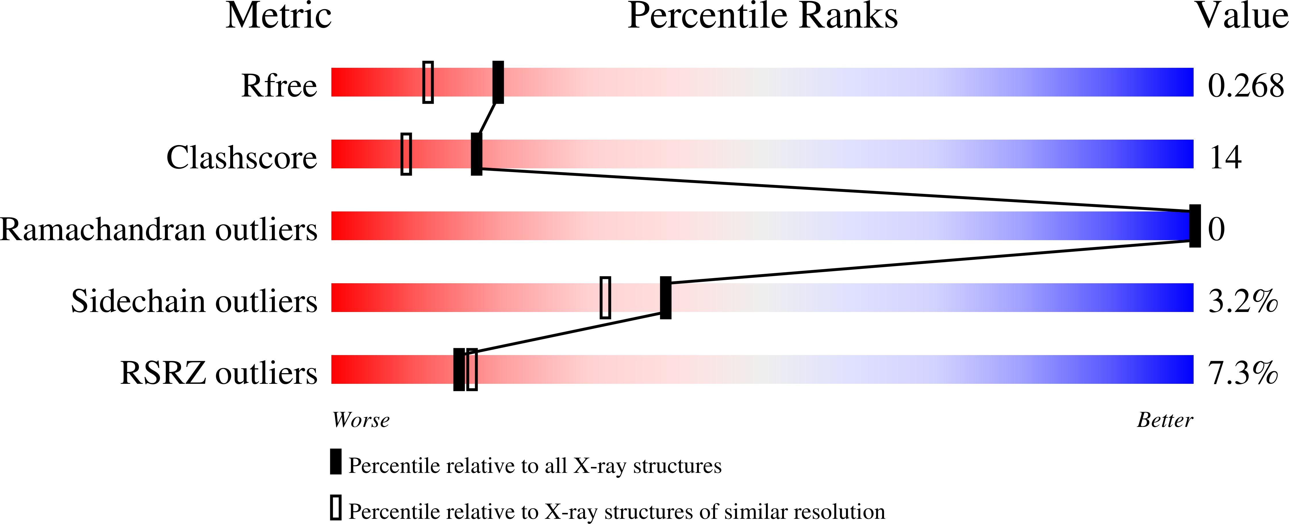
    2ZFH

  • PubMed Abstract: 

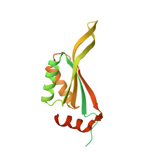
    The structure of human brain CutA1 (HsCutA1) has been determined using diffraction data to 2.05 A resolution. HsCutA1 has been implicated in the anchoring of acetylcholinesterase in neuronal cell membranes, while its bacterial homologue Escherichia coli CutA1 is involved in copper tolerance. Additionally, the structure of HsCutA1 bears similarity to that of the signal transduction protein PII, which is involved in regulation of nitrogen metabolism. Although several crystal structures of CutA1 from various sources with different rotation angles and degrees of interaction between trimer interfaces have been reported, the specific functional role of CutA1 is still unclear. In this study, the X-ray structure of HsCutA1 was determined in space group P2(1)2(1)2(1), with unit-cell parameters a = 68.69, b = 88.84, c = 125.33 A and six molecules per asymmetric unit. HsCutA1 is a trimeric molecule with intertwined antiparallel beta-strands; each subunit has a molecular weight of 14.6 kDa and contains 135 amino-acid residues. In order to obtain clues to the possible function of HsCutA1, its crystal structure was compared with those of other CutA1 and PII proteins.


  • Organizational Affiliation

    Protein Structure Analysis Team, RIKEN SPring-8 Center, Harima Institute, 1-1-1 Kouto, Sayo-cho, Sayo-gun, Hyogo 679-5148, Japan. bagautdi@spring8.or.jp


Macromolecules
Find similar proteins by:  (by identity cutoff)  |  3D Structure
Entity ID: 1
MoleculeChains Sequence LengthOrganismDetailsImage
CutA
A, B, C, D, E
A, B, C, D, E, F
179Homo sapiensMutation(s): 0 
Gene Names: CUTAACHAPC6orf82
UniProt & NIH Common Fund Data Resources
Find proteins for O60888 (Homo sapiens)
Explore O60888 
Go to UniProtKB:  O60888
PHAROS:  O60888
GTEx:  ENSG00000112514 
Entity Groups  
Sequence Clusters30% Identity50% Identity70% Identity90% Identity95% Identity100% Identity
UniProt GroupO60888
Sequence Annotations
Expand
  • Reference Sequence
Experimental Data & Validation

Experimental Data

  • Method: X-RAY DIFFRACTION
  • Resolution: 2.05 Å
  • R-Value Free: 0.265 
  • R-Value Work: 0.221 
  • R-Value Observed: 0.221 
  • Space Group: P 21 21 21
Unit Cell:
Length ( Å )Angle ( ˚ )
a = 68.693α = 90
b = 88.844β = 90
c = 125.334γ = 90
Software Package:
Software NamePurpose
HKL-2000data collection
MOLREPphasing
CNSrefinement
HKL-2000data reduction
SCALEPACKdata scaling

Structure Validation

View Full Validation Report


Entry History 

Deposition Data

Revision History  (Full details and data files)

  • Version 1.0: 2008-01-22
    Type: Initial release
  • Version 1.1: 2011-07-13
    Changes: Version format compliance
  • Version 1.2: 2012-04-18
    Changes: Database references, Derived calculations
  • Version 1.3: 2023-11-01
    Changes: Data collection, Database references, Refinement description