Novel inhibitor for prolyl tripeptidyl aminopeptidase from Porphyromonas gingivalis and details of substrate-recognition mechanism
Xu, Y., Nakajima, Y., Ito, K., Zheng, H., Oyama, H., Heiser, U., Hoffmann, T., Gartner, U.T., Demuth, H.U., Yoshimoto, T.(2008) J Mol Biol 375: 708-719
- PubMed: 18042490
- DOI: https://doi.org/10.1016/j.jmb.2007.09.077
- Primary Citation of Related Structures:
2EEP, 2Z3W, 2Z3Z - PubMed Abstract:
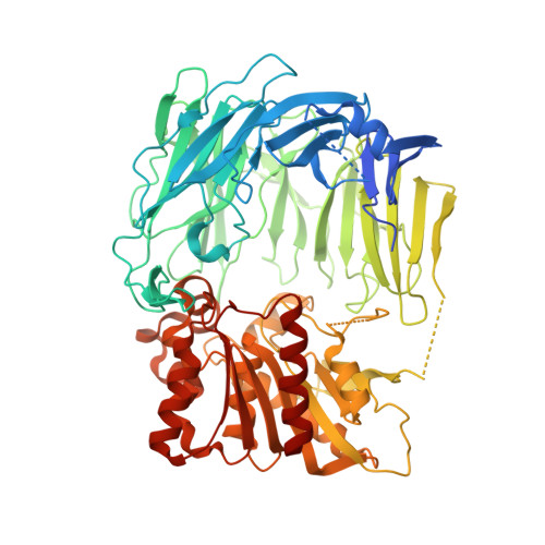
A new inhibitor, H-Ala-Ile-pyrrolidin-2-yl boronic acid, was developed as an inhibitor against prolyl tripeptidyl aminopeptidase with a K(i) value of 88.1 nM. The structure of the prolyl tripeptidyl aminopeptidase complexed with the inhibitor (enzyme-inhibitor complex) was determined at 2.2 A resolution. The inhibitor was bound to the active site through a covalent bond between Ser603 and the boron atom of the inhibitor. This structure should closely mimic the structure of the reaction intermediate between the enzyme and substrate. We previously proposed that two glutamate residues, Glu205 and Glu636, are involved in the recognition of substrates. In order to clarify the function of these glutamate residues in substrate recognition, three mutant enzymes, E205A, E205Q, and E636A were generated by site-directed mutagenesis. The E205A mutant was expressed as an inclusion body. The E205Q mutant was expressed in soluble form, but no activity was detected. Here, the structures of the E636A mutant and its complex with the inhibitor were determined. The inhibitor was located at almost the same position as in the wild-type enzyme-inhibitor complex. The amino group of the inhibitor interacted with Glu205 and the main-chain carbonyl group of Gln203. In addition, a water molecule in the place of Glu636 of the wild-type enzyme interacted with the amino group of the inhibitor. This water molecule was located near the position of Glu636 in the wild-type and formed a hydrogen bond with Gln203. The k(cat)/K(M) values of the E636A mutant toward the two substrates used were smaller than those of the wild-type by two orders of magnitude. The K(i) value of our inhibitor for the E636A mutant was 48.8 microM, which was 554-fold higher than that against the wild-type enzyme. Consequently, it was concluded that Glu205 and Glu636 are significant residues for the N-terminal recognition of a substrate.
Organizational Affiliation:
Graduate School of Biomedical Sciences, Nagasaki University, 1-14 Bunkyo-machi, Nagasaki 852-8521, Japan.