2X6Y

Tailspike protein mutant D339A of E.coli bacteriophage HK620 in complex with hexasaccharide


Experimental Data Snapshot

  • Method: X-RAY DIFFRACTION
  • Resolution: 1.35 Å
  • R-Value Free: 0.171 
  • R-Value Work: 0.132 
  • R-Value Observed: 0.134 

wwPDB Validation   3D Report Full Report


This is version 2.1 of the entry. See complete history


Literature

Single Amino Acid Exchange in Bacteriophage Hk620 Tailspike Protein Results in Thousand-Fold Increase of its Oligosaccharide Affinity.

Broeker, N.K.Gohlke, U.Muller, J.J.Uetrecht, C.Heinemann, U.Seckler, R.Barbirz, S.

(2013) Glycobiology 23: 59

  • DOI: https://doi.org/10.1093/glycob/cws126
  • Primary Citation of Related Structures:  
    2X6W, 2X6X, 2X6Y, 2X85, 4AVZ

  • PubMed Abstract: 

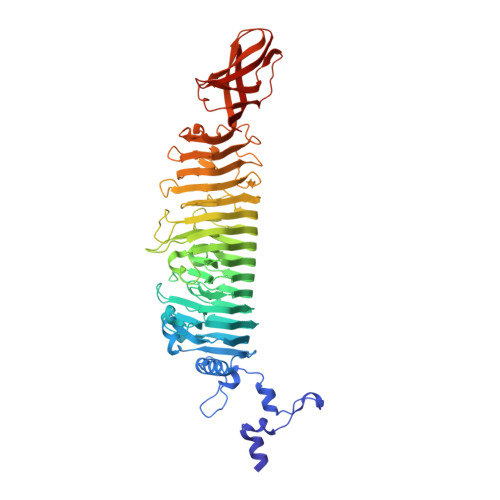
    Bacteriophage HK620 recognizes and cleaves the O-antigen polysaccharide of Escherichia coli serogroup O18A1 with its tailspike protein (TSP). HK620TSP binds hexasaccharide fragments with low affinity, but single amino acid exchanges generated a set of high-affinity mutants with submicromolar dissociation constants. Isothermal titration calorimetry showed that only small amounts of heat were released upon complex formation via a large number of direct and solvent-mediated hydrogen bonds between carbohydrate and protein. At room temperature, association was both enthalpy- and entropy-driven emphasizing major solvent rearrangements upon complex formation. Crystal structure analysis showed identical protein and sugar conformers in the TSP complexes regardless of their hexasaccharide affinity. Only in one case, a TSP mutant bound a different hexasaccharide conformer. The extended sugar binding site could be dissected in two regions: first, a hydrophobic pocket at the reducing end with minor affinity contributions. Access to this site could be blocked by a single aspartate to asparagine exchange without major loss in hexasaccharide affinity. Second, a region where the specific exchange of glutamate for glutamine created a site for an additional water molecule. Side-chain rearrangements upon sugar binding led to desolvation and additional hydrogen bonding which define this region of the binding site as the high-affinity scaffold.


  • Organizational Affiliation

    Physikalische Biochemie, Universität Potsdam, Germany.


Macromolecules
Find similar proteins by:  (by identity cutoff)  |  3D Structure
Entity ID: 1
MoleculeChains Sequence LengthOrganismDetailsImage
TAIL SPIKE PROTEIN600Enterobacteria phage HK620Mutation(s): 1 
UniProt
Find proteins for Q9AYY6 (Enterobacteria phage HK620)
Explore Q9AYY6 
Go to UniProtKB:  Q9AYY6
Entity Groups  
Sequence Clusters30% Identity50% Identity70% Identity90% Identity95% Identity100% Identity
UniProt GroupQ9AYY6
Sequence Annotations
Expand
  • Reference Sequence
Oligosaccharides

Help

Entity ID: 2
MoleculeChains Length2D Diagram Glycosylation3D Interactions
alpha-L-rhamnopyranose-(1-6)-alpha-D-glucopyranose-(1-4)-[2-acetamido-2-deoxy-beta-D-glucopyranose-(1-3)]alpha-D-galactopyranose-(1-3)-[alpha-D-glucopyranose-(1-6)]2-acetamido-2-deoxy-beta-D-glucopyranose
B
6N/A
Glycosylation Resources
GlyTouCan:  G22623XU
GlyCosmos:  G22623XU
Experimental Data & Validation

Experimental Data

  • Method: X-RAY DIFFRACTION
  • Resolution: 1.35 Å
  • R-Value Free: 0.171 
  • R-Value Work: 0.132 
  • R-Value Observed: 0.134 
  • Space Group: P 3 2 1
Unit Cell:
Length ( Å )Angle ( ˚ )
a = 74.336α = 90
b = 74.336β = 90
c = 174.977γ = 120
Software Package:
Software NamePurpose
REFMACrefinement
XDSdata reduction
XSCALEdata scaling
PHASERphasing

Structure Validation

View Full Validation Report


Entry History 

Deposition Data

Revision History  (Full details and data files)

  • Version 1.0: 2011-03-02
    Type: Initial release
  • Version 1.1: 2014-02-05
    Changes: Database references, Version format compliance
  • Version 1.2: 2019-01-30
    Changes: Data collection, Experimental preparation, Other
  • Version 1.3: 2019-02-06
    Changes: Data collection, Experimental preparation
  • Version 2.0: 2020-07-29
    Type: Remediation
    Reason: Carbohydrate remediation
    Changes: Atomic model, Data collection, Derived calculations, Other, Structure summary
  • Version 2.1: 2023-12-20
    Changes: Data collection, Database references, Refinement description, Structure summary