2VWC

STRUCTURE OF THE HSP90 INHIBITOR MACBECIN BOUND TO THE N-TERMINUS OF YEAST HSP90.


Experimental Data Snapshot

  • Method: X-RAY DIFFRACTION
  • Resolution: 2.40 Å
  • R-Value Free: 0.262 
  • R-Value Work: 0.195 
  • R-Value Observed: 0.198 

wwPDB Validation   3D Report Full Report


Ligand Structure Quality Assessment 


This is version 1.2 of the entry. See complete history


Literature

Molecular Characterisation of Macbecin as an Hsp90 Inhibitor

Martin, C.J.Gaisser, S.Challis, I.R.Carletti, I.Wilkinson, B.Gregory, M.Prodromou, C.Roe, S.M.Pearl, L.H.Boyd, S.M.Zhang, M.Q.

(2008) J Med Chem 51: 2853

  • DOI: https://doi.org/10.1021/jm701558c
  • Primary Citation of Related Structures:  
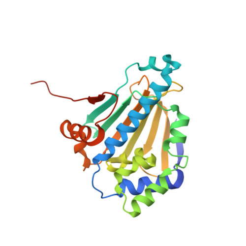
    2VWC

  • PubMed Abstract: 

    Macbecin compares favorably to geldanamycin as an Hsp90 inhibitor, being more soluble, stable, more potently inhibiting ATPase activity (IC50 = 2 microM) and binding with higher affinity (Kd = 0.24 microM). Structural studies reveal significant differences in their Hsp90 binding characteristics, and macbecin-induced tumor cell growth inhibition is accompanied by characteristic degradation of Hsp90 client proteins. Macbecin significantly reduced tumor growth rates (minimum T/C: 32%) in a DU145 murine xenograft. Macbecin thus represents an attractive lead for further optimization.


  • Organizational Affiliation

    Biotica Technology Limited, Chesterford Research Park, Essex CB10 1XL, U.K. christine.martin@biotica.com


Macromolecules
Find similar proteins by:  (by identity cutoff)  |  3D Structure
Entity ID: 1
MoleculeChains Sequence LengthOrganismDetailsImage
ATP-DEPENDENT MOLECULAR CHAPERONE HSP82219Saccharomyces cerevisiaeMutation(s): 0 
UniProt
Find proteins for P02829 (Saccharomyces cerevisiae (strain ATCC 204508 / S288c))
Explore P02829 
Go to UniProtKB:  P02829
Entity Groups  
Sequence Clusters30% Identity50% Identity70% Identity90% Identity95% Identity100% Identity
UniProt GroupP02829
Sequence Annotations
Expand
  • Reference Sequence
Small Molecules
Binding Affinity Annotations 
IDSourceBinding Affinity
BC2 Binding MOAD:  2VWC Kd: 240 (nM) from 1 assay(s)
PDBBind:  2VWC Kd: 240 (nM) from 1 assay(s)
Experimental Data & Validation

Experimental Data

  • Method: X-RAY DIFFRACTION
  • Resolution: 2.40 Å
  • R-Value Free: 0.262 
  • R-Value Work: 0.195 
  • R-Value Observed: 0.198 
  • Space Group: P 43 2 2
Unit Cell:
Length ( Å )Angle ( ˚ )
a = 73.805α = 90
b = 73.805β = 90
c = 110.667γ = 90
Software Package:
Software NamePurpose
REFMACrefinement
MOSFLMdata reduction
SCALAdata scaling
PHASERphasing

Structure Validation

View Full Validation Report


Ligand Structure Quality Assessment 


Entry History 

Deposition Data

Revision History  (Full details and data files)

  • Version 1.0: 2008-07-01
    Type: Initial release
  • Version 1.1: 2011-07-13
    Changes: Advisory, Version format compliance
  • Version 1.2: 2023-12-13
    Changes: Data collection, Database references, Other, Refinement description