2VT8

Structure of a conserved dimerisation domain within Fbox7 and PI31


Experimental Data Snapshot

  • Method: X-RAY DIFFRACTION
  • Resolution: 2.60 Å
  • R-Value Free: 0.256 
  • R-Value Work: 0.203 
  • R-Value Observed: 0.206 

wwPDB Validation   3D Report Full Report


This is version 1.2 of the entry. See complete history


Literature

Structure of a Conserved Dimerization Domain within the F-Box Protein Fbxo7 and the Pi31 Proteasome Inhibitor.

Kirk, R.J.Laman, H.Knowles, P.P.Murray-Rust, J.Lomonosov, M.Meziane, E.K.McDonald, N.Q.

(2008) J Biol Chem 283: 22325

  • DOI: https://doi.org/10.1074/jbc.M709900200
  • Primary Citation of Related Structures:  
    2VT8

  • PubMed Abstract: 

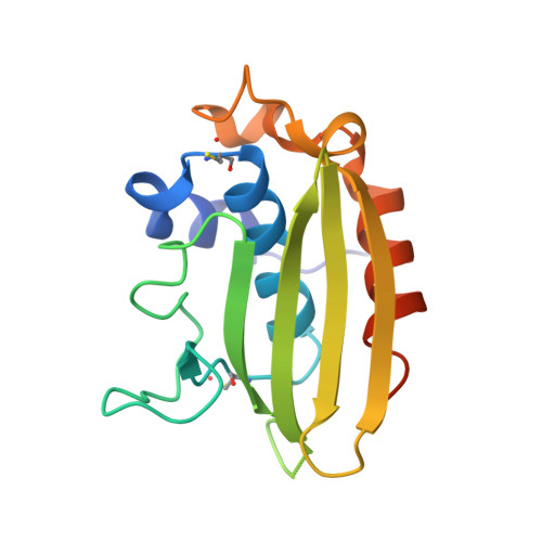
    F-box proteins are the substrate-recognition components of the Skp1-Cul1-F box protein (SCF) E3 ubiquitin ligases. Here we report a structural relationship between Fbxo7, a component of the SCF(Fbxo7) E3 ligase, and the proteasome inhibitor PI31. SCF(Fbxo7) is known to catalyze the ubiquitination of hepatoma-up-regulated protein (HURP) and the inhibitor of apoptosis (IAP) protein but also functions as an activator of cyclin D-Cdk6 complexes. We identify PI31 as an Fbxo7.Skp1 binding partner and show that this interaction requires an N-terminal domain present in both proteins that we term the FP (Fbxo7/PI31) domain. The crystal structure of the PI31 FP domain reveals a novel alpha/beta-fold. Biophysical and mutational analyses are used to map regions of the PI31 FP domain mediating homodimerization and required for heterodimerization with Fbxo7.Skp1. Equivalent mutations in Fbxo7 ablate interaction with PI31 and also block Fbxo7 homodimerization. Knockdown of Fbxo7 does not affect PI31 levels arguing against PI31 being a substrate for SCF(Fbxo7). We present a model for FP domain-mediated dimerization of SCF(Fbxo7) and PI31.


  • Organizational Affiliation

    Structural Biology Laboratory, London Research Institute, Cancer Research UK, 44 Lincoln's Inn Fields, London WC2A 3PX, United Kingdom.


Macromolecules
Find similar proteins by:  (by identity cutoff)  |  3D Structure
Entity ID: 1
MoleculeChains Sequence LengthOrganismDetailsImage
PROTEASOME INHIBITOR PI31 SUBUNIT
A, B
153Homo sapiensMutation(s): 1 
UniProt & NIH Common Fund Data Resources
Find proteins for Q92530 (Homo sapiens)
Explore Q92530 
Go to UniProtKB:  Q92530
PHAROS:  Q92530
GTEx:  ENSG00000125818 
Entity Groups  
Sequence Clusters30% Identity50% Identity70% Identity90% Identity95% Identity100% Identity
UniProt GroupQ92530
Sequence Annotations
Expand
  • Reference Sequence
Small Molecules
Modified Residues  1 Unique
IDChains TypeFormula2D DiagramParent
CSO
Query on CSO
A, B
L-PEPTIDE LINKINGC3 H7 N O3 SCYS
Experimental Data & Validation

Experimental Data

  • Method: X-RAY DIFFRACTION
  • Resolution: 2.60 Å
  • R-Value Free: 0.256 
  • R-Value Work: 0.203 
  • R-Value Observed: 0.206 
  • Space Group: C 1 2 1
Unit Cell:
Length ( Å )Angle ( ˚ )
a = 109.498α = 90
b = 45.212β = 111.54
c = 67.279γ = 90
Software Package:
Software NamePurpose
REFMACrefinement
MOSFLMdata reduction
SCALAdata scaling
MOLREPphasing

Structure Validation

View Full Validation Report


Entry History 

Deposition Data

Revision History  (Full details and data files)

  • Version 1.0: 2008-05-20
    Type: Initial release
  • Version 1.1: 2012-06-06
    Changes: Database references, Derived calculations, Non-polymer description, Other, Structure summary, Version format compliance
  • Version 1.2: 2018-03-07
    Changes: Database references