The Three-Dimensional Structure of the Cytoplasmic Domains of Epsf from the Type 2 Secretion System of Vibrio Cholerae.
Abendroth, J., Mitchell, D.D., Korotkov, K.V., Johnson, T.L., Kreger, A., Sandkvist, M., Hol, W.G.(2009) J Struct Biol 166: 303
- PubMed: 19324092
- DOI: https://doi.org/10.1016/j.jsb.2009.03.009
- Primary Citation of Related Structures:
2VMA, 2VMB, 3C1Q - PubMed Abstract:
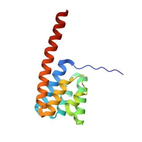
The type 2 secretion system (T2SS), a multi-protein machinery that spans both the inner and the outer membranes of Gram-negative bacteria, is used for the secretion of several critically important proteins across the outer membrane. Here we report the crystal structure of the N-terminal cytoplasmic domain of EpsF, an inner membrane spanning T2SS protein from Vibrio cholerae. This domain consists of a bundle of six anti-parallel helices and adopts a fold that has not been described before. The long C-terminal helix alpha6 protrudes from the body of the domain and most likely continues as the first transmembrane helix of EpsF. Two N-terminal EpsF domains form a tight dimer with a conserved interface, suggesting that the observed dimer occurs in the T2SS of many bacteria. Two calcium binding sites are present in the dimer interface with ligands provided for each site by both subunits. Based on this new structure, sequence comparisons of EpsF homologs and localization studies of GFP fused with EpsF, we propose that the second cytoplasmic domain of EpsF adopts a similar fold as the first cytoplasmic domain and that full-length EpsF, and its T2SS homologs, have a three-transmembrane helix topology.
Organizational Affiliation:
Department of Biochemistry, Biomolecular Structure Center, University of Washington, 1959 Pacific Ave. NE, Box 357742, Seattle, WA 98195, USA.