2V95

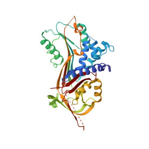
Structure of Corticosteroid-Binding Globulin in complex with Cortisol


Experimental Data Snapshot

  • Method: X-RAY DIFFRACTION
  • Resolution: 1.93 Å
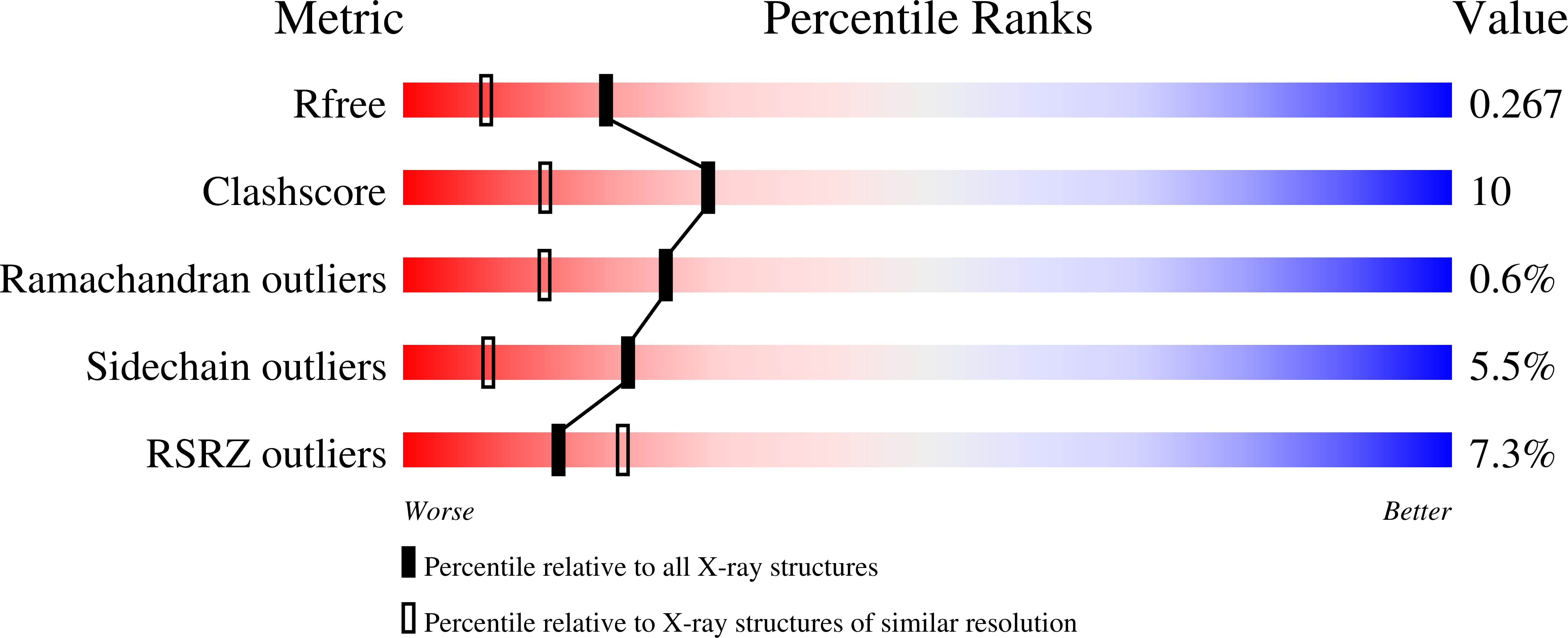
  • R-Value Free: 0.264 
  • R-Value Work: 0.210 
  • R-Value Observed: 0.214 

wwPDB Validation   3D Report Full Report


Ligand Structure Quality Assessment 


This is version 1.2 of the entry. See complete history


Literature

Corticosteroid-Binding Globulin: Structural Basis for Steroid Transport and Proteinase-Triggered Release

Klieber, M.A.Underhill, C.Hammond, G.L.Muller, Y.A.

(2007) J Biol Chem 282: 29594

  • DOI: https://doi.org/10.1074/jbc.M705014200
  • Primary Citation of Related Structures:  
    2V95

  • PubMed Abstract: 

    Corticosteroid-binding globulin (CBG) is a serine proteinase inhibitor (serpin) family member that transports glucocorticoids in blood and regulates their access to target cells. The 1.9A crystal structure of rat CBG shows that its steroid-binding site resembles the thyroxin-binding site in the related serpin, thyroxin-binding globulin, and mutagenesis studies have confirmed the contributions of key residues that constitute the steroid-binding pocket. Unlike thyroxin-bound thyroxin-binding globulin, the cortisol-bound CBG displays an "active" serpin conformation with the proteinase-sensitive, reactive center loop (RCL) fully expelled from the regulatory beta-sheet A. Moreover, the CBG structure allows us to predict that complete insertion of the proteolytically cleaved RCL into the serpin fold occurs in concert with a displacement and unwinding of helix D that would disrupt the steroid-binding site. This allosteric coupling between RCL positioning and occupancy of the CBG steroid-binding site, which resembles the ligand (glycosamino-glycan)-dependent activation of the thrombin inhibitory serpins heparin cofactor II and anti-thrombin RCLs, ensures both optimal recognition of CBG by target proteinases and efficient release of steroid to sites of action.


  • Organizational Affiliation

    Lehrstuhl für Biotechnik, Department of Biology, Friedrich-Alexander-University Erlangen-Nuremberg, D-91052 Erlangen, Germany.


Macromolecules
Find similar proteins by:  (by identity cutoff)  |  3D Structure
Entity ID: 1
MoleculeChains Sequence LengthOrganismDetailsImage
CORTICOSTEROID-BINDING GLOBULIN371Rattus norvegicusMutation(s): 2 
UniProt
Find proteins for P31211 (Rattus norvegicus)
Explore P31211 
Go to UniProtKB:  P31211
Entity Groups  
Sequence Clusters30% Identity50% Identity70% Identity90% Identity95% Identity100% Identity
UniProt GroupP31211
Sequence Annotations
Expand
  • Reference Sequence
Small Molecules
Ligands 1 Unique
IDChains Name / Formula / InChI Key2D Diagram3D Interactions
HCY
Query on HCY

Download Ideal Coordinates CCD File 
B [auth A](11alpha,14beta)-11,17,21-trihydroxypregn-4-ene-3,20-dione
C21 H30 O5
JYGXADMDTFJGBT-VWUMJDOOSA-N
Binding Affinity Annotations 
IDSourceBinding Affinity
PDN PDBBind:  2V95 Kd: 5 (nM) from 1 assay(s)
Experimental Data & Validation

Experimental Data

  • Method: X-RAY DIFFRACTION
  • Resolution: 1.93 Å
  • R-Value Free: 0.264 
  • R-Value Work: 0.210 
  • R-Value Observed: 0.214 
  • Space Group: C 1 2 1
Unit Cell:
Length ( Å )Angle ( ˚ )
a = 122.11α = 90
b = 54.25β = 97.16
c = 61.04γ = 90
Software Package:
Software NamePurpose
REFMACrefinement
XDSdata reduction
XSCALEdata scaling
AMoREphasing

Structure Validation

View Full Validation Report


Ligand Structure Quality Assessment 


Entry History 

Deposition Data

Revision History  (Full details and data files)

  • Version 1.0: 2007-09-04
    Type: Initial release
  • Version 1.1: 2015-12-02
    Changes: Derived calculations, Other, Refinement description, Structure summary, Version format compliance
  • Version 1.2: 2023-12-13
    Changes: Data collection, Database references, Derived calculations, Other, Refinement description