2V6W

tRNASer acceptor stem: Conformation and hydration of a microhelix in a crystal structure at 1.8 Angstrom resolution


Experimental Data Snapshot

  • Method: X-RAY DIFFRACTION
  • Resolution: 1.80 Å
  • R-Value Work: 0.154 

wwPDB Validation   3D Report Full Report


This is version 1.4 of the entry. See complete history


Literature

Trnaser Acceptor Stem: Conformation and Hydration of a Microhelix in a Crystal Structure at 1.8 A Resolution.

Forster, C.Brauer, A.B.E.Brode, S.Furste, J.P.Betzel, C.Erdmann, V.A.

(2007) Acta Crystallogr D Biol Crystallogr 63: 1154

  • DOI: https://doi.org/10.1107/S0907444907045271
  • Primary Citation of Related Structures:  
    2V6W

  • PubMed Abstract: 

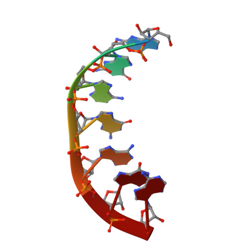
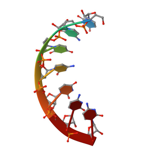
    The crystal structure of a serine-specific tRNA acceptor-stem microhelix, the binding site for the seryl-tRNA synthetase, was solved by X-ray analysis. This seven-base-pair tRNA(Ser) microhelix forms endless rows of helices in the crystal lattice, with two helices stacking 'head-to-head' onto each other, resulting in an intermolecular guanosine stacking of the first purine nucleotides at the 5'-strands of the tRNA(Ser) microhelices. A network of 75 water loci could be associated with each RNA duplex. Unusual local geometric backbone parameters could be detected in the region of the G4 phosphate located in the 5'-strand of the helix, which lead to a ;kink' in this region and to an irregularly bent helix. The role of the specific hydration pattern and of the irregular conformation of the tRNA(Ser) acceptor-stem helix is discussed and summarized.


  • Organizational Affiliation

    Institute of Chemistry and Biochemistry, Free University Berlin, Thielallee 63, 14195 Berlin, Germany.


Macromolecules

Find similar nucleic acids by:  Sequence   |   3D Structure  

Entity ID: 1
MoleculeChains LengthOrganismImage
5'-R(*GP*GP*AP*GP*AP*GP*AP)-3'7Escherichia coli
Sequence Annotations
Expand
  • Reference Sequence

Find similar nucleic acids by:  Sequence   |   3D Structure  

Entity ID: 2
MoleculeChains LengthOrganismImage
5'-R(*UP*CP*UP*CP*UP*CP*CP)-3'7Escherichia coli
Sequence Annotations
Expand
  • Reference Sequence
Experimental Data & Validation

Experimental Data

  • Method: X-RAY DIFFRACTION
  • Resolution: 1.80 Å
  • R-Value Work: 0.154 
  • Space Group: C 1 2 1
Unit Cell:
Length ( Å )Angle ( ˚ )
a = 36.1α = 90
b = 38.92β = 110.61
c = 30.79γ = 90
Software Package:
Software NamePurpose
REFMACrefinement

Structure Validation

View Full Validation Report


Entry History 

Deposition Data

Revision History  (Full details and data files)

  • Version 1.0: 2007-11-06
    Type: Initial release
  • Version 1.1: 2011-05-08
    Changes: Version format compliance
  • Version 1.2: 2011-07-13
    Changes: Version format compliance
  • Version 1.3: 2019-05-08
    Changes: Data collection, Experimental preparation, Other
  • Version 1.4: 2024-05-08
    Changes: Data collection, Database references, Other