2V6H

Crystal structure of the C1 domain of cardiac myosin binding protein-C


Experimental Data Snapshot

  • Method: X-RAY DIFFRACTION
  • Resolution: 1.55 Å
  • R-Value Free: 0.241 
  • R-Value Observed: 0.184 

wwPDB Validation   3D Report Full Report


This is version 1.4 of the entry. See complete history


Literature

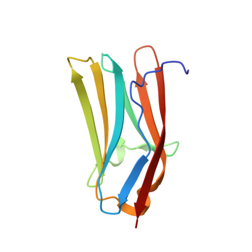
Crystal structure of the C1 domain of cardiac myosin binding protein-C: implications for hypertrophic cardiomyopathy.

Govada, L.Carpenter, L.da Fonseca, P.C.Helliwell, J.R.Rizkallah, P.Flashman, E.Chayen, N.E.Redwood, C.Squire, J.M.

(2008) J Mol Biol 378: 387-397

  • DOI: https://doi.org/10.1016/j.jmb.2008.02.044
  • Primary Citation of Related Structures:  
    2V6H

  • PubMed Abstract: 

    C-protein is a major component of skeletal and cardiac muscle thick filaments. Mutations in the gene encoding cardiac C-protein [cardiac myosin binding protein-C (cMyBP-C)] are one of the principal causes of hypertrophic cardiomyopathy. cMyBP-C is a string of globular domains including eight immunoglobulin-like and three fibronectin-like domains termed C0-C10. It binds to myosin and titin, and probably to actin, and may have both a structural and a regulatory role in muscle function. To help to understand the pathology of the known mutations, we have solved the structure of the immunoglobulin-like C1 domain of MyBP-C by X-ray crystallography to a resolution of 1.55 A. Mutations associated with hypertrophic cardiomyopathy are clustered at one end towards the C-terminus, close to the important C1C2 linker, where they alter the structural integrity of this region and its interactions.


  • Organizational Affiliation

    Biomolecular Medicine Department, SORA Division, Imperial College London, London SW7 2AZ, UK.


Macromolecules
Find similar proteins by:  (by identity cutoff)  |  3D Structure
Entity ID: 1
MoleculeChains Sequence LengthOrganismDetailsImage
MYOSIN-BINDING PROTEIN C, CARDIAC-TYPE108Homo sapiensMutation(s): 0 
UniProt & NIH Common Fund Data Resources
Find proteins for Q14896 (Homo sapiens)
Explore Q14896 
Go to UniProtKB:  Q14896
PHAROS:  Q14896
GTEx:  ENSG00000134571 
Entity Groups  
Sequence Clusters30% Identity50% Identity70% Identity90% Identity95% Identity100% Identity
UniProt GroupQ14896
Sequence Annotations
Expand
  • Reference Sequence
Experimental Data & Validation

Experimental Data

  • Method: X-RAY DIFFRACTION
  • Resolution: 1.55 Å
  • R-Value Free: 0.241 
  • R-Value Observed: 0.184 
  • Space Group: I 41
Unit Cell:
Length ( Å )Angle ( ˚ )
a = 48.848α = 90
b = 48.848β = 90
c = 95.132γ = 90
Software Package:
Software NamePurpose
HKL-2000data reduction
SOLVEphasing
RESOLVEphasing
CNSphasing
REFMACphasing
SHELXL-97phasing
Cootmodel building
SHELXL-97refinement

Structure Validation

View Full Validation Report


Entry History 

Deposition Data

Revision History  (Full details and data files)

  • Version 1.0: 2008-07-22
    Type: Initial release
  • Version 1.1: 2011-05-08
    Changes: Version format compliance
  • Version 1.2: 2011-07-13
    Changes: Version format compliance
  • Version 1.3: 2017-08-23
    Changes: Database references, Refinement description
  • Version 1.4: 2019-05-08
    Changes: Data collection, Experimental preparation