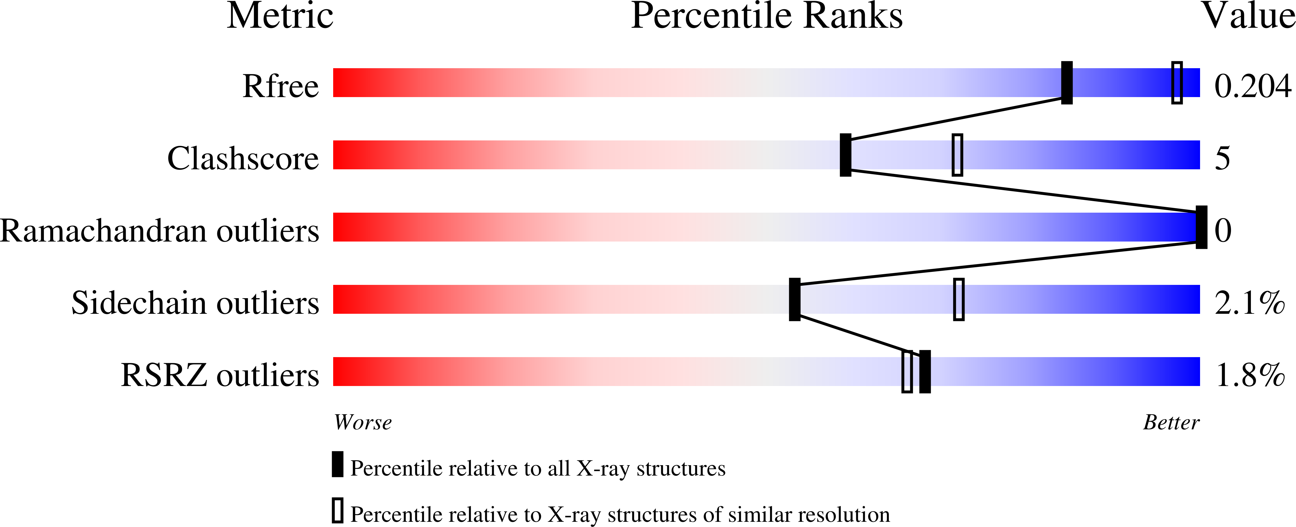
2V3A

Crystal structure of rubredoxin reductase from Pseudomonas aeruginosa.


Experimental Data Snapshot

  • Method: X-RAY DIFFRACTION
  • Resolution: 2.40 Å
  • R-Value Free: 0.202 
  • R-Value Work: 0.166 
  • R-Value Observed: 0.168 

wwPDB Validation   3D Report Full Report


This is version 1.2 of the entry. See complete history


Literature

Crystal Structure of the Electron Transfer Complex Rubredoxin - Rubredoxin Reductase from Pseudomonas Aeruginosa.

Hagelueken, G.Wiehlmann, L.Adams, T.M.Kolmar, H.Heinz, D.W.Tuemmler, B.Schubert, W.-D.

(2007) Proc Natl Acad Sci U S A 104: 12276

  • DOI: https://doi.org/10.1073/pnas.0702919104
  • Primary Citation of Related Structures:  
    2V3A, 2V3B

  • PubMed Abstract: 

    Crude oil spills represent a major ecological threat because of the chemical inertness of the constituent n-alkanes. The Gram-negative bacterium Pseudomonas aeruginosa is one of the few bacterial species able to metabolize such compounds. Three chromosomal genes, rubB, rubA1, and rubA2 coding for an NAD(P)H:rubredoxin reductase (RdxR) and two rubredoxins (Rdxs) are indispensable for this ability. They constitute an electron transport (ET) pathway that shuttles reducing equivalents from carbon metabolism to the membrane-bound alkane hydroxylases AlkB1 and AlkB2. The RdxR-Rdx system also is crucial as part of the oxidative stress response in archaea or anaerobic bacteria. The redox couple has been analyzed in detail as a model system for ET processes. We have solved the structure of RdxR of P. aeruginosa both alone and in complex with Rdx, without the need for cross-linking, and both structures were refined at 2.40- and 2.45-A resolution, respectively. RdxR consists of two cofactor-binding domains and a C-terminal domain essential for the specific recognition of Rdx. Only a small number of direct interactions govern mutual recognition of RdxR and Rdx, corroborating the transient nature of the complex. The shortest distance between the redox centers is observed to be 6.2 A.


  • Organizational Affiliation

    Molecular Host-Pathogen Interactions, Division of Structural Biology, Helmholtz Centre for Infection Research, Inhoffenstrasse 7, D-38124 Braunschweig, Germany.


Macromolecules
Find similar proteins by:  (by identity cutoff)  |  3D Structure
Entity ID: 1
MoleculeChains Sequence LengthOrganismDetailsImage
RUBREDOXIN REDUCTASE384Pseudomonas aeruginosa PAO1Mutation(s): 0 
UniProt
Find proteins for Q9HTK9 (Pseudomonas aeruginosa (strain ATCC 15692 / DSM 22644 / CIP 104116 / JCM 14847 / LMG 12228 / 1C / PRS 101 / PAO1))
Explore Q9HTK9 
Go to UniProtKB:  Q9HTK9
Entity Groups  
Sequence Clusters30% Identity50% Identity70% Identity90% Identity95% Identity100% Identity
UniProt GroupQ9HTK9
Sequence Annotations
Expand
  • Reference Sequence
Small Molecules
Ligands 3 Unique
IDChains Name / Formula / InChI Key2D Diagram3D Interactions
FAD
Query on FAD

Download Ideal Coordinates CCD File 
J [auth A]FLAVIN-ADENINE DINUCLEOTIDE
C27 H33 N9 O15 P2
VWWQXMAJTJZDQX-UYBVJOGSSA-N
PEG
Query on PEG

Download Ideal Coordinates CCD File 
B [auth A]
C [auth A]
D [auth A]
E [auth A]
F [auth A]
B [auth A],
C [auth A],
D [auth A],
E [auth A],
F [auth A],
G [auth A]
DI(HYDROXYETHYL)ETHER
C4 H10 O3
MTHSVFCYNBDYFN-UHFFFAOYSA-N
CL
Query on CL

Download Ideal Coordinates CCD File 
H [auth A],
I [auth A]
CHLORIDE ION
Cl
VEXZGXHMUGYJMC-UHFFFAOYSA-M
Experimental Data & Validation

Experimental Data

  • Method: X-RAY DIFFRACTION
  • Resolution: 2.40 Å
  • R-Value Free: 0.202 
  • R-Value Work: 0.166 
  • R-Value Observed: 0.168 
  • Space Group: P 61 2 2
Unit Cell:
Length ( Å )Angle ( ˚ )
a = 119.602α = 90
b = 119.602β = 90
c = 158.064γ = 120
Software Package:
Software NamePurpose
REFMACrefinement
HKL-2000data reduction
SCALEPACKdata scaling
PHASERphasing

Structure Validation

View Full Validation Report


Entry History 

Deposition Data

Revision History  (Full details and data files)

  • Version 1.0: 2007-08-14
    Type: Initial release
  • Version 1.1: 2011-07-13
    Changes: Advisory, Version format compliance
  • Version 1.2: 2023-12-13
    Changes: Data collection, Database references, Other, Refinement description