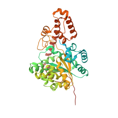
Catalytic mechanism of the tryptophan activation reaction revealed by crystal structures of human tryptophanyl-tRNA synthetase in different enzymatic states
Shen, N., Zhou, M., Yang, B., Yu, Y., Dong, X., Ding, J.(2008) Nucleic Acids Res 36: 1288-1299
- PubMed: 18180246
- DOI: https://doi.org/10.1093/nar/gkm1153
- Primary Citation of Related Structures:
2QUH, 2QUI, 2QUJ, 2QUK - PubMed Abstract:
Human tryptophanyl-tRNA synthetase (hTrpRS) differs from its bacterial counterpart at several key positions of the catalytic active site and has an extra N-terminal domain, implying possibly a different catalytic mechanism. We report here the crystal structures of hTrpRS in complexes with Trp, tryptophanamide and ATP and tryptophanyl-AMP, respectively, which represent three different enzymatic states of the Trp activation reaction. Analyses of these structures reveal the molecular basis of the mechanisms of the substrate recognition and the activation reaction. The dimeric hTrpRS is structurally and functionally asymmetric with half-of-the-sites reactivity. Recognition of Trp is by an induced-fit mechanism involving conformational change of the AIDQ motif that creates a perfect pocket for the binding and activation of Trp and causes coupled movements of the N-terminal and C-terminal domains. The KMSAS loop appears to have an inherent flexibility and the binding of ATP stabilizes it in a closed conformation that secures the position of ATP for catalysis. Our structural data indicate that the catalytic mechanism of the Trp activation reaction by hTrpRS involves more moderate conformational changes of the structural elements at the active site to recognize and bind the substrates, which is more complex and fine-tuned than that of bacterial TrpRS.
Organizational Affiliation:
State Key Laboratory of Molecular Biology, Institute of Biochemistry and Cell Biology, Shanghai Institutes for Biological Sciences, Chinese Academy of Sciences, and Graduate School of Chinese Academy of Sciences, 320 Yue-Yang Road, Shanghai 200031, China.