2QT6

Crystal Structure Determination of a Blue Laccase from Lentinus Tigrinus


Experimental Data Snapshot

  • Method: X-RAY DIFFRACTION
  • Resolution: 1.50 Å
  • R-Value Free: 0.182 
  • R-Value Work: 0.150 
  • R-Value Observed: 0.150 

wwPDB Validation   3D Report Full Report


Ligand Structure Quality Assessment 


This is version 3.1 of the entry. See complete history


Literature

Crystal structure of a blue laccase from Lentinus tigrinus: evidences for intermediates in the molecular oxygen reductive splitting by multicopper oxidases.

Ferraroni, M.Myasoedova, N.M.Schmatchenko, V.Leontievsky, A.A.Golovleva, L.A.Scozzafava, A.Briganti, F.

(2007) BMC Struct Biol 7: 60-60

  • DOI: https://doi.org/10.1186/1472-6807-7-60
  • Primary Citation of Related Structures:  
    2QT6

  • PubMed Abstract: 

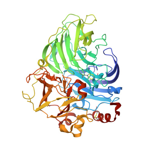
    Laccases belong to multicopper oxidases, a widespread class of enzymes implicated in many oxidative functions in pathogenesis, immunogenesis and morphogenesis of organisms and in the metabolic turnover of complex organic substances. They catalyze the coupling between the four one-electron oxidations of a broad range of substrates with the four-electron reduction of dioxygen to water. These catalytic processes are made possible by the contemporaneous presence of at least four copper ion sites, classified according to their spectroscopic properties: one type 1 (T1) site where the electrons from the reducing substrates are accepted, one type 2 (T2), and a coupled binuclear type 3 pair (T3) which are assembled in a T2/T3 trinuclear cluster where the electrons are transferred to perform the O2 reduction to H2O. The structure of a laccase from the white-rot fungus Lentinus (Panus) tigrinus, a glycoenzyme involved in lignin biodegradation, was solved at 1.5 A. It reveals a asymmetric unit containing two laccase molecules (A and B). The progressive reduction of the copper ions centers obtained by the long-term exposure of the crystals to the high-intensity X-ray synchrotron beam radiation under aerobic conditions and high pH allowed us to detect two sequential intermediates in the molecular oxygen reduction pathway: the "peroxide" and the "native" intermediates, previously hypothesized through spectroscopic, kinetic and molecular mechanics studies. Specifically the electron-density maps revealed the presence of an end-on bridging, micro-eta 1:eta 1 peroxide ion between the two T3 coppers in molecule B, result of a two-electrons reduction, whereas in molecule A an oxo ion bridging the three coppers of the T2/T3 cluster (micro3-oxo bridge) together with an hydroxide ion externally bridging the two T3 copper ions, products of the four-electrons reduction of molecular oxygen, were best modelled. This is the first structure of a multicopper oxidase which allowed the detection of two intermediates in the molecular oxygen reduction and splitting. The observed features allow to positively substantiate an accurate mechanism of dioxygen reduction catalyzed by multicopper oxidases providing general insights into the reductive cleavage of the O-O bonds, a leading problem in many areas of biology.


  • Organizational Affiliation

    Department of Chemistry University of Florence, Via della Lastruccia 3, Sesto Fiorentino, 50019 Florence, Italy. mferraroni@unifi.it


Macromolecules
Find similar proteins by:  (by identity cutoff)  |  3D Structure
Entity ID: 1
MoleculeChains Sequence LengthOrganismDetailsImage
Laccase
A, B
498Lentinus tigrinusMutation(s): 0 
EC: 1.10.3.2
UniProt
Find proteins for Q5EBY5 (Lentinus tigrinus)
Explore Q5EBY5 
Go to UniProtKB:  Q5EBY5
Entity Groups  
Sequence Clusters30% Identity50% Identity70% Identity90% Identity95% Identity100% Identity
UniProt GroupQ5EBY5
Sequence Annotations
Expand
  • Reference Sequence
Oligosaccharides

Help

Entity ID: 2
MoleculeChains Length2D Diagram Glycosylation3D Interactions
beta-D-mannopyranose-(1-6)-beta-D-mannopyranose-(1-6)-[beta-D-mannopyranose-(1-3)]beta-D-mannopyranose-(1-4)-2-acetamido-2-deoxy-beta-D-glucopyranose-(1-4)-2-acetamido-2-deoxy-beta-D-glucopyranose
C
6N-Glycosylation
Glycosylation Resources
GlyTouCan:  G52684JK
GlyCosmos:  G52684JK
GlyGen:  G52684JK
Entity ID: 3
MoleculeChains Length2D Diagram Glycosylation3D Interactions
beta-D-mannopyranose-(1-3)-beta-D-mannopyranose-(1-6)-[beta-D-mannopyranose-(1-3)]beta-D-mannopyranose-(1-4)-2-acetamido-2-deoxy-beta-D-glucopyranose-(1-4)-2-acetamido-2-deoxy-beta-D-glucopyranose
D
6N-Glycosylation
Glycosylation Resources
GlyTouCan:  G67849DJ
GlyCosmos:  G67849DJ
GlyGen:  G67849DJ
Entity ID: 4
MoleculeChains Length2D Diagram Glycosylation3D Interactions
2-acetamido-2-deoxy-beta-D-glucopyranose-(1-4)-2-acetamido-2-deoxy-beta-D-glucopyranoseE [auth F],
F [auth G]
2N-Glycosylation
Glycosylation Resources
GlyTouCan:  G42666HT
GlyCosmos:  G42666HT
GlyGen:  G42666HT
Small Molecules
Ligands 8 Unique
IDChains Name / Formula / InChI Key2D Diagram3D Interactions
NAG
Query on NAG

Download Ideal Coordinates CCD File 
K [auth A]2-acetamido-2-deoxy-beta-D-glucopyranose
C8 H15 N O6
OVRNDRQMDRJTHS-FMDGEEDCSA-N
MAN
Query on MAN

Download Ideal Coordinates CCD File 
BA [auth B],
N [auth A]
alpha-D-mannopyranose
C6 H12 O6
WQZGKKKJIJFFOK-PQMKYFCFSA-N
TLA
Query on TLA

Download Ideal Coordinates CCD File 
L [auth A]L(+)-TARTARIC ACID
C4 H6 O6
FEWJPZIEWOKRBE-JCYAYHJZSA-N
GOL
Query on GOL

Download Ideal Coordinates CCD File 
AA [auth B],
M [auth A]
GLYCEROL
C3 H8 O3
PEDCQBHIVMGVHV-UHFFFAOYSA-N
CU
Query on CU

Download Ideal Coordinates CCD File 
G [auth A]
H [auth A]
I [auth A]
J [auth A]
V [auth B]
G [auth A],
H [auth A],
I [auth A],
J [auth A],
V [auth B],
W [auth B],
X [auth B],
Y [auth B]
COPPER (II) ION
Cu
JPVYNHNXODAKFH-UHFFFAOYSA-N
CA
Query on CA

Download Ideal Coordinates CCD File 
CA [auth B]
O [auth A]
P [auth A]
Q [auth A]
R [auth A]
CA [auth B],
O [auth A],
P [auth A],
Q [auth A],
R [auth A],
S [auth A]
CALCIUM ION
Ca
BHPQYMZQTOCNFJ-UHFFFAOYSA-N
CL
Query on CL

Download Ideal Coordinates CCD File 
DA [auth B],
T [auth A],
U [auth A]
CHLORIDE ION
Cl
VEXZGXHMUGYJMC-UHFFFAOYSA-M
PER
Query on PER

Download Ideal Coordinates CCD File 
Z [auth B]PEROXIDE ION
O2
ANAIPYUSIMHBEL-UHFFFAOYSA-N
Experimental Data & Validation

Experimental Data

  • Method: X-RAY DIFFRACTION
  • Resolution: 1.50 Å
  • R-Value Free: 0.182 
  • R-Value Work: 0.150 
  • R-Value Observed: 0.150 
  • Space Group: P 1 21 1
Unit Cell:
Length ( Å )Angle ( ˚ )
a = 54.218α = 90
b = 111.613β = 97.75
c = 97.086γ = 90
Software Package:
Software NamePurpose
DENZOdata reduction
SCALEPACKdata scaling
MOLREPphasing
REFMACrefinement
PDB_EXTRACTdata extraction
MAR345dtbdata collection

Structure Validation

View Full Validation Report


Ligand Structure Quality Assessment 


Entry History 

Deposition Data

Revision History  (Full details and data files)

  • Version 1.0: 2007-11-20
    Type: Initial release
  • Version 1.1: 2011-07-13
    Changes: Non-polymer description, Version format compliance
  • Version 1.2: 2017-10-25
    Changes: Advisory, Refinement description
  • Version 2.0: 2020-07-29
    Type: Remediation
    Reason: Carbohydrate remediation
    Changes: Advisory, Atomic model, Data collection, Derived calculations, Non-polymer description, Structure summary
  • Version 3.0: 2022-04-20
    Changes: Advisory, Atomic model, Data collection, Database references, Derived calculations, Source and taxonomy, Structure summary
  • Version 3.1: 2023-06-21
    Changes: Derived calculations