2QQW

Crystal structure of a cell-wall invertase (D23A) from Arabidopsis thaliana in complex with sucrose


Experimental Data Snapshot

  • Method: X-RAY DIFFRACTION
  • Resolution: 2.80 Å
  • R-Value Free: 0.252 
  • R-Value Work: 0.188 
  • R-Value Observed: 0.192 

wwPDB Validation   3D Report Full Report


Ligand Structure Quality Assessment 


This is version 2.2 of the entry. See complete history


Literature

Crystal structures of Arabidopsis thaliana cell-wall invertase mutants in complex with sucrose.

Lammens, W.Le Roy, K.Van Laere, A.Rabijns, A.Van den Ende, W.

(2008) J Mol Biol 377: 378-385

  • DOI: https://doi.org/10.1016/j.jmb.2007.12.074
  • Primary Citation of Related Structures:  
    2QQU, 2QQV, 2QQW

  • PubMed Abstract: 

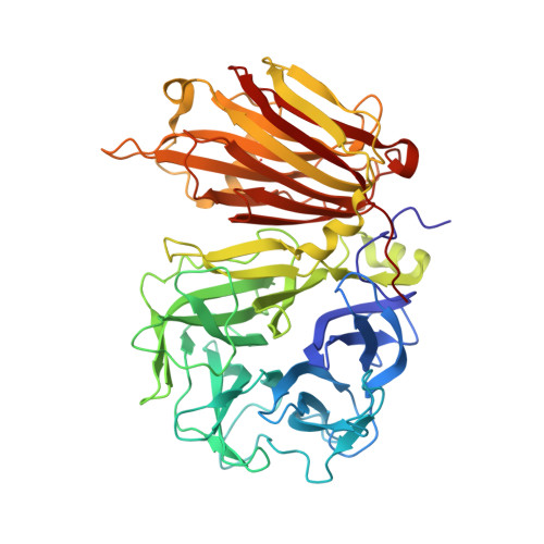
    In plants, cell-wall invertases fulfil important roles in carbohydrate partitioning, growth, development and crop yield. In this study, we report on different X-ray crystal structures of Arabidopsis thaliana cell-wall invertase 1 (AtcwINV1) mutants with sucrose. These structures reveal a detailed view of sucrose binding in the active site of the wild-type AtcwINV1. Compared to related enzyme-sucrose complexes, important differences in the orientation of the glucose subunit could be observed. The structure of the E203Q AtcwINV1 mutant showed a complete new binding modus, whereas the D23A, E203A and D239A structures most likely represent the productive binding modus. Together with a hydrophobic zone formed by the conserved W20, W47 and W82, the residues N22, D23, R148, E203, D149 and D239 are necessary to create the ideal sucrose-binding pocket. D239 can interact directly with the glucose moiety of sucrose, whereas K242 has an indirect role in substrate stabilization. Most probably, K242 keeps D239 in a favourable position upon substrate binding. Unravelling the exact position of sucrose in plant cell-wall invertases is a necessary step towards the rational design of superior invertases to further increase crop yield and biomass production.


  • Organizational Affiliation

    Laboratorium voor Moleculaire Plantenfysiologie, Faculteit Wetenschappen, Departement Biologie, K.U. Leuven, Kasteelpark Arenberg 31, bus 2434, B-3001 Heverlee, Belgium.


Macromolecules
Find similar proteins by:  (by identity cutoff)  |  3D Structure
Entity ID: 1
MoleculeChains Sequence LengthOrganismDetailsImage
Beta-fructofuranosidase537Arabidopsis thalianaMutation(s): 1 
Gene Names: ATBFRUCT1
EC: 3.2.1.26
UniProt
Find proteins for Q43866 (Arabidopsis thaliana)
Explore Q43866 
Go to UniProtKB:  Q43866
Entity Groups  
Sequence Clusters30% Identity50% Identity70% Identity90% Identity95% Identity100% Identity
UniProt GroupQ43866
Sequence Annotations
Expand
  • Reference Sequence
Oligosaccharides

Help

Entity ID: 2
MoleculeChains Length2D Diagram Glycosylation3D Interactions
alpha-D-mannopyranose-(1-2)-alpha-D-mannopyranose-(1-6)-[alpha-D-mannopyranose-(1-3)]alpha-D-mannopyranose-(1-4)-2-acetamido-2-deoxy-beta-D-glucopyranose-(1-4)-2-acetamido-2-deoxy-beta-D-glucopyranose
B
6N-Glycosylation
Glycosylation Resources
GlyTouCan:  G08161XL
GlyCosmos:  G08161XL
GlyGen:  G08161XL
Entity ID: 3
MoleculeChains Length2D Diagram Glycosylation3D Interactions
2-acetamido-2-deoxy-beta-D-glucopyranose-(1-4)-2-acetamido-2-deoxy-beta-D-glucopyranose
C
2N-Glycosylation
Glycosylation Resources
GlyTouCan:  G42666HT
GlyCosmos:  G42666HT
GlyGen:  G42666HT
Entity ID: 4
MoleculeChains Length2D Diagram Glycosylation3D Interactions
beta-D-fructofuranose-(2-1)-alpha-D-glucopyranose
D
2N/A
Glycosylation Resources
GlyTouCan:  G05551OP
GlyCosmos:  G05551OP
Biologically Interesting Molecules (External Reference) 1 Unique
Experimental Data & Validation

Experimental Data

  • Method: X-RAY DIFFRACTION
  • Resolution: 2.80 Å
  • R-Value Free: 0.252 
  • R-Value Work: 0.188 
  • R-Value Observed: 0.192 
  • Space Group: P 32
Unit Cell:
Length ( Å )Angle ( ˚ )
a = 105.877α = 90
b = 105.877β = 90
c = 50.479γ = 120
Software Package:
Software NamePurpose
DENZOdata reduction
SCALEPACKdata scaling
MOLREPphasing
REFMACrefinement
PDB_EXTRACTdata extraction
MAR345dtbdata collection

Structure Validation

View Full Validation Report


Ligand Structure Quality Assessment 


Entry History 

Deposition Data

Revision History  (Full details and data files)

  • Version 1.0: 2008-04-22
    Type: Initial release
  • Version 1.1: 2011-07-13
    Changes: Non-polymer description, Version format compliance
  • Version 1.2: 2017-10-25
    Changes: Refinement description
  • Version 2.0: 2020-07-29
    Type: Remediation
    Reason: Carbohydrate remediation
    Changes: Advisory, Atomic model, Data collection, Derived calculations, Non-polymer description, Structure summary
  • Version 2.1: 2021-10-20
    Changes: Database references, Structure summary
  • Version 2.2: 2023-08-30
    Changes: Data collection, Refinement description