2QEH

Crystal Structure of Anopheles gambiae D7r4-serotonin complex


Experimental Data Snapshot

  • Method: X-RAY DIFFRACTION
  • Resolution: 2.10 Å
  • R-Value Free: 0.238 
  • R-Value Work: 0.193 
  • R-Value Observed: 0.196 

wwPDB Validation   3D Report Full Report


Ligand Structure Quality Assessment 


This is version 1.2 of the entry. See complete history


Literature

The Crystal Structure of D7r4, a Salivary Biogenic Amine-binding Protein from the Malaria Mosquito Anopheles gambiae

Mans, B.J.Calvo, E.Ribeiro, J.M.Andersen, J.F.

(2007) J Biol Chem 282: 36626-36633

  • DOI: https://doi.org/10.1074/jbc.M706410200
  • Primary Citation of Related Structures:  
    2PQL, 2QEB, 2QEH, 2QEO, 2QEV

  • PubMed Abstract: 

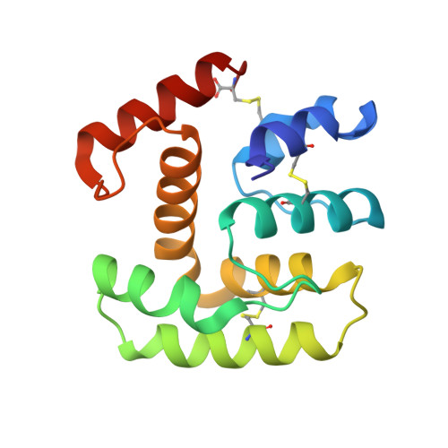
    The D7-related (D7r) proteins of the malaria vector Anopheles gambiae have been shown to bind the biogenic amines serotonin, norepinephrine, and histamine with high affinity. One member of the group (D7r1 or hamadarin) has also been shown to have an anticoagulant/antikinin activity. To understand the mechanistic details of its antihemostatic/anti-inflammatory effects, we have determined the crystal structure of one member of this group, D7r4, along with the structures of ligand complexes with serotonin, tryptamine, histamine, and norepinephrine. The D7 fold consists of an arrangement of eight alpha-helices stabilized by three disulfide bonds. The structure is similar to those of the arthropod odorant-binding proteins, a relationship that had been predicted based on sequence comparisons. Although odorant-binding proteins commonly have six alpha-helices, D7r4 has eight, resulting in significantly different positioning and structure of the ligand binding pocket. The pocket itself is lined by hydrophobic side chains along with polar and charged groups oriented to form hydrogen bonds with the aliphatic amino group and with groups on the aromatic portions of the ligands. These structures, along with accompanying mutagenesis studies, have allowed us to identify critical residues for biogenic amine binding and to predict which members of the large D7 protein family found in blood-feeding nematocerous Diptera will function as biogenic amine-binding proteins.


  • Organizational Affiliation

    Laboratory of Malaria and Vector Research, NIAID, National Institutes of Health, Rockville, Maryland 20852, USA.


Macromolecules
Find similar proteins by:  (by identity cutoff)  |  3D Structure
Entity ID: 1
MoleculeChains Sequence LengthOrganismDetailsImage
D7R4 Protein145Anopheles gambiaeMutation(s): 0 
Gene Names: D7r4
UniProt
Find proteins for Q7PNF2 (Anopheles gambiae)
Explore Q7PNF2 
Go to UniProtKB:  Q7PNF2
Entity Groups  
Sequence Clusters30% Identity50% Identity70% Identity90% Identity95% Identity100% Identity
UniProt GroupQ7PNF2
Sequence Annotations
Expand
  • Reference Sequence
Small Molecules
Ligands 1 Unique
IDChains Name / Formula / InChI Key2D Diagram3D Interactions
SRO
Query on SRO

Download Ideal Coordinates CCD File 
B [auth A]SEROTONIN
C10 H12 N2 O
QZAYGJVTTNCVMB-UHFFFAOYSA-N
Binding Affinity Annotations 
IDSourceBinding Affinity
SRO Binding MOAD:  2QEH Kd: 2.4 (nM) from 1 assay(s)
Experimental Data & Validation

Experimental Data

  • Method: X-RAY DIFFRACTION
  • Resolution: 2.10 Å
  • R-Value Free: 0.238 
  • R-Value Work: 0.193 
  • R-Value Observed: 0.196 
  • Space Group: P 43
Unit Cell:
Length ( Å )Angle ( ˚ )
a = 63.358α = 90
b = 63.358β = 90
c = 42.617γ = 90
Software Package:
Software NamePurpose
DENZOdata reduction
SCALEPACKdata scaling
REFMACrefinement
PDB_EXTRACTdata extraction
EPICS-baseddata collection
HKL-3000data reduction

Structure Validation

View Full Validation Report


Ligand Structure Quality Assessment 


Entry History 

Deposition Data

Revision History  (Full details and data files)

  • Version 1.0: 2007-10-09
    Type: Initial release
  • Version 1.1: 2011-07-13
    Changes: Advisory, Version format compliance
  • Version 1.2: 2017-10-18
    Changes: Refinement description