2QCO

Crystal structure of the transcriptional regulator CmeR from Campylobacter jejuni


Experimental Data Snapshot

  • Method: X-RAY DIFFRACTION
  • Resolution: 2.25 Å
  • R-Value Free: 0.243 
  • R-Value Work: 0.219 
  • R-Value Observed: 0.230 

wwPDB Validation   3D Report Full Report


This is version 1.3 of the entry. See complete history


Literature

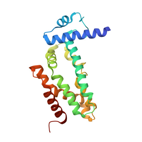
Crystal structure of the transcriptional regulator CmeR from Campylobacter jejuni.

Gu, R.Su, C.C.Shi, F.Li, M.McDermott, G.Zhang, Q.Yu, E.W.

(2007) J Mol Biol 372: 583-593

  • DOI: https://doi.org/10.1016/j.jmb.2007.06.072
  • Primary Citation of Related Structures:  
    2QCO

  • PubMed Abstract: 

    The CmeABC multidrug efflux pump, which belongs to the resistance-nodulation-division (RND) family, recognizes and extrudes a broad range of antimicrobial agents and is essential for Campylobacter jejuni colonization of the animal intestinal tract by mediating the efflux of bile acids. The expression of CmeABC is controlled by the transcriptional regulator CmeR, whose open reading frame is located immediately upstream of the cmeABC operon. To understand the structural basis of CmeR regulation, we have determined the crystal structure of CmeR to 2.2 A resolution, revealing a dimeric two-domain molecule with an entirely helical architecture similar to members of the TetR family of transcriptional regulators. Unlike the rest of the TetR regulators, CmeR has a large center-to-center distance (54 A) between two N termini of the dimer, and a large flexible ligand-binding pocket in the C-terminal domain. Each monomer forms a 20 A long tunnel-like cavity in the ligand-binding domain of CmeR and is occupied by a fortuitous ligand that is identified as glycerol. The binding of glycerol to CmeR induces a conformational state that is incompatible with target DNA. As glycerol has a chemical structure similar to that of potential ligands of CmeR, the structure obtained mimics the induced form of CmeR. These findings reveal novel structural features of a TetR family regulator, and provide new insight into the mechanisms of ligand binding and CmeR regulation.


  • Organizational Affiliation

    Department of Physics and Astronomy, Iowa State University, Ames, IA 50011, USA.


Macromolecules
Find similar proteins by:  (by identity cutoff)  |  3D Structure
Entity ID: 1
MoleculeChains Sequence LengthOrganismDetailsImage
CmeR210Campylobacter jejuniMutation(s): 0 
Gene Names: cmeR
UniProt
Find proteins for Q7B8P6 (Campylobacter jejuni)
Explore Q7B8P6 
Go to UniProtKB:  Q7B8P6
Entity Groups  
Sequence Clusters30% Identity50% Identity70% Identity90% Identity95% Identity100% Identity
UniProt GroupQ7B8P6
Sequence Annotations
Expand
  • Reference Sequence
Small Molecules
Ligands 1 Unique
IDChains Name / Formula / InChI Key2D Diagram3D Interactions
GOL
Query on GOL

Download Ideal Coordinates CCD File 
B [auth A]GLYCEROL
C3 H8 O3
PEDCQBHIVMGVHV-UHFFFAOYSA-N
Experimental Data & Validation

Experimental Data

  • Method: X-RAY DIFFRACTION
  • Resolution: 2.25 Å
  • R-Value Free: 0.243 
  • R-Value Work: 0.219 
  • R-Value Observed: 0.230 
  • Space Group: P 21 21 2
Unit Cell:
Length ( Å )Angle ( ˚ )
a = 93.125α = 90
b = 37.587β = 90
c = 57.789γ = 90
Software Package:
Software NamePurpose
HKL-2000data collection
SnBphasing
CNSrefinement
HKL-2000data reduction
HKL-2000data scaling

Structure Validation

View Full Validation Report


Entry History 

Deposition Data

Revision History  (Full details and data files)

  • Version 1.0: 2008-04-15
    Type: Initial release
  • Version 1.1: 2011-07-13
    Changes: Derived calculations, Version format compliance
  • Version 1.2: 2017-10-18
    Changes: Refinement description
  • Version 1.3: 2024-02-21
    Changes: Data collection, Database references, Derived calculations