2Q2C

Crystal structures of the arginine-, lysine-, histidine-binding protein ArtJ from the thermophilic bacterium Geobacillus stearothermophilus


Experimental Data Snapshot

  • Method: X-RAY DIFFRACTION
  • Resolution: 2.35 Å
  • R-Value Free: 0.333 
  • R-Value Work: 0.268 
  • R-Value Observed: 0.270 

wwPDB Validation   3D Report Full Report


Ligand Structure Quality Assessment 


This is version 1.3 of the entry. See complete history


Literature

Crystal structures and mutational analysis of the arginine-, lysine-, histidine-binding protein ArtJ from Geobacillus stearothermophilus. Implications for interactions of ArtJ with its cognate ATP-binding cassette transporter, Art(MP)2

Vahedi-Faridi, A.Eckey, V.Scheffel, F.Alings, C.Landmesser, H.Schneider, E.Saenger, W.

(2008) J Mol Biol 375: 448-459

  • DOI: https://doi.org/10.1016/j.jmb.2007.10.049
  • Primary Citation of Related Structures:  
    2PVU, 2Q2A, 2Q2C

  • PubMed Abstract: 

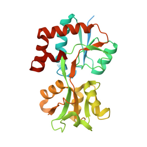
    ArtJ is the substrate-binding component (receptor) of the ATP-binding cassette (ABC) transport system ArtJ-(MP)(2) from the thermophilic bacterium Geobacillus stearothermophilus that is specific for arginine, lysine, and histidine. The highest affinity is found for arginine (K(d)=0.039(+/-0.014) microM), while the affinities for lysine and histidine are about tenfold lower. We have determined the X-ray structures of ArtJ liganded with each of these substrates at resolutions of 1.79 A (arginine), 1.79 A (lysine), and 2.35 A (histidine), respectively. As found for other solute receptors, the polypeptide chain is folded into two distinct domains (lobes) connected by a hinge. The interface between the lobes forms the substrate-binding pocket whose geometry is well preserved in all three ArtJ/amino acid complexes. Structure-derived mutational analyses indicated the crucial role of a region in the carboxy-terminal lobe of ArtJ in contacting the transport pore Art(MP)(2) and revealed the functional importance of Gln132 and Trp68. While variant Gln132Leu exhibited lower binding affinity for arginine but no binding of lysine and histidine, the variant Trp68Leu had lost binding activity for all three substrates. The results are discussed in comparison with known structures of homologous proteins from mesophilic bacteria.


  • Organizational Affiliation

    Institut für Chemie und Biochemie/Kristallographie, Freie Universitaet Berlin, Takustr. 6, 14195 Berlin, Germany.


Macromolecules
Find similar proteins by:  (by identity cutoff)  |  3D Structure
Entity ID: 1
MoleculeChains Sequence LengthOrganismDetailsImage
ArtJ
A, B, C, D
272Geobacillus stearothermophilusMutation(s): 0 
UniProt
Find proteins for D0VWX8 (Geobacillus stearothermophilus)
Explore D0VWX8 
Go to UniProtKB:  D0VWX8
Entity Groups  
Sequence Clusters30% Identity50% Identity70% Identity90% Identity95% Identity100% Identity
UniProt GroupD0VWX8
Sequence Annotations
Expand
  • Reference Sequence
Small Molecules
Ligands 3 Unique
IDChains Name / Formula / InChI Key2D Diagram3D Interactions
HIS
Query on HIS

Download Ideal Coordinates CCD File 
F [auth A],
I [auth B],
K [auth C],
N [auth D]
HISTIDINE
C6 H10 N3 O2
HNDVDQJCIGZPNO-YFKPBYRVSA-O
SO4
Query on SO4

Download Ideal Coordinates CCD File 
E [auth A],
G [auth B],
H [auth B],
J [auth C],
M [auth D]
SULFATE ION
O4 S
QAOWNCQODCNURD-UHFFFAOYSA-L
GOL
Query on GOL

Download Ideal Coordinates CCD File 
L [auth C],
O [auth D]
GLYCEROL
C3 H8 O3
PEDCQBHIVMGVHV-UHFFFAOYSA-N
Binding Affinity Annotations 
IDSourceBinding Affinity
HIS Binding MOAD:  2Q2C Kd: 420 (nM) from 1 assay(s)
PDBBind:  2Q2C Kd: 420 (nM) from 1 assay(s)
Experimental Data & Validation

Experimental Data

  • Method: X-RAY DIFFRACTION
  • Resolution: 2.35 Å
  • R-Value Free: 0.333 
  • R-Value Work: 0.268 
  • R-Value Observed: 0.270 
  • Space Group: P 1 21 1
Unit Cell:
Length ( Å )Angle ( ˚ )
a = 67.07α = 90
b = 102.2β = 95.36
c = 70.535γ = 90
Software Package:
Software NamePurpose
DENZOdata reduction
SCALEPACKdata scaling
REFMACrefinement
PDB_EXTRACTdata extraction
ADSCdata collection
HKL-2000data reduction

Structure Validation

View Full Validation Report


Ligand Structure Quality Assessment 


Entry History 

Deposition Data

Revision History  (Full details and data files)

  • Version 1.0: 2008-01-15
    Type: Initial release
  • Version 1.1: 2011-07-13
    Changes: Non-polymer description, Version format compliance
  • Version 1.2: 2017-10-18
    Changes: Refinement description
  • Version 1.3: 2024-02-21
    Changes: Data collection, Database references, Derived calculations