2PYW

Structure of A. thaliana 5-methylthioribose kinase in complex with ADP and MTR


Experimental Data Snapshot

  • Method: X-RAY DIFFRACTION
  • Resolution: 1.90 Å
  • R-Value Free: 0.197 
  • R-Value Work: 0.163 
  • R-Value Observed: 0.165 

wwPDB Validation   3D Report Full Report


Ligand Structure Quality Assessment 


This is version 2.1 of the entry. See complete history


Literature

Structure of A. thaliana 5-methylthioribose kinase in complex with ADP and MTR reveals a more occluded active site than its bacterial homolog

Ku, S.Y.Cornell, K.A.Howell, P.L.

(2007) BMC Struct Biol 7: 70-70

  • DOI: https://doi.org/10.1186/1472-6807-7-70
  • Primary Citation of Related Structures:  
    2PYW

  • PubMed Abstract: 

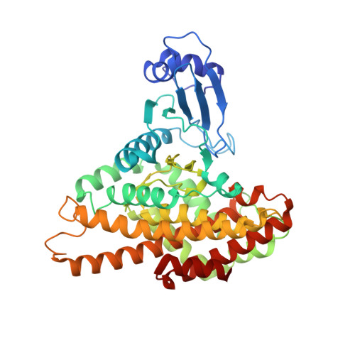
    Metabolic variations exist between the methionine salvage pathway of humans and a number of plants and microbial pathogens. 5-Methylthioribose (MTR) kinase is a key enzyme required for methionine salvage in plants and many bacteria. The absence of a mammalian homolog suggests that MTR kinase is a good target for the design of specific herbicides or antibiotics. The structure of Arabidopsis thaliana MTR kinase co-crystallized with ATPgammaS and MTR has been determined at 1.9 A resolution. The structure is similar to B. subtilis MTR kinase and has the same protein kinase fold observed in other evolutionarily related protein kinase-like phosphotransferases. The active site is comparable between the two enzymes with the DXE-motif coordinating the nucleotide-Mg, the D238 of the HGD catalytic loop polarizing the MTR O1 oxygen, and the RR-motif interacting with the substrate MTR. Unlike its bacterial homolog, however, the Gly-rich loop (G-loop) of A. thaliana MTR kinase has an extended conformation, which shields most of the active site from solvent, a feature that resembles eukaryotic protein kinases more than the bacterial enzyme. The G- and W-loops of A. thaliana and B. subtilis MTR kinase adopt different conformations despite high sequence similarity. The ATPgammaS analog was hydrolyzed during the co-crystallization procedure, resulting in ADP in the active site. This suggests that the A. thaliana enzyme, like its bacterial homolog, may have significant ATPase activity in the absence of MTR. The structure of A. thaliana MTR kinase provides a template for structure-based design of agrochemicals, particularly herbicides whose effectiveness could be regulated by nutrient levels. Features of the MTR binding site offer an opportunity for a simple organic salt of an MTR analog to specifically inhibit MTR kinase.


  • Organizational Affiliation

    Program in Molecular Structure and Function, Research Institute, Hospital for Sick Children, 555 University Avenue, Toronto, Ontario, M5G 1X8, CANADA. syku@sickkids.ca


Macromolecules
Find similar proteins by:  (by identity cutoff)  |  3D Structure
Entity ID: 1
MoleculeChains Sequence LengthOrganismDetailsImage
Uncharacterized protein
A, B
420Arabidopsis thalianaMutation(s): 0 
Gene Names: F10F5.1
EC: 2.7.1.100
UniProt
Find proteins for Q9C6D2 (Arabidopsis thaliana)
Explore Q9C6D2 
Go to UniProtKB:  Q9C6D2
Entity Groups  
Sequence Clusters30% Identity50% Identity70% Identity90% Identity95% Identity100% Identity
UniProt GroupQ9C6D2
Sequence Annotations
Expand
  • Reference Sequence
Small Molecules
Ligands 5 Unique
IDChains Name / Formula / InChI Key2D Diagram3D Interactions
ADP
Query on ADP

Download Ideal Coordinates CCD File 
H [auth A],
N [auth B]
ADENOSINE-5'-DIPHOSPHATE
C10 H15 N5 O10 P2
XTWYTFMLZFPYCI-KQYNXXCUSA-N
SR1
Query on SR1

Download Ideal Coordinates CCD File 
G [auth A],
M [auth B]
5-S-methyl-5-thio-alpha-D-ribofuranose
C6 H12 O4 S
OLVVOVIFTBSBBH-KAZBKCHUSA-N
EDO
Query on EDO

Download Ideal Coordinates CCD File 
I [auth A],
O [auth B],
P [auth B],
Q [auth B],
R [auth B]
1,2-ETHANEDIOL
C2 H6 O2
LYCAIKOWRPUZTN-UHFFFAOYSA-N
CL
Query on CL

Download Ideal Coordinates CCD File 
C [auth A],
J [auth B]
CHLORIDE ION
Cl
VEXZGXHMUGYJMC-UHFFFAOYSA-M
MG
Query on MG

Download Ideal Coordinates CCD File 
D [auth A],
E [auth A],
F [auth A],
K [auth B],
L [auth B]
MAGNESIUM ION
Mg
JLVVSXFLKOJNIY-UHFFFAOYSA-N
Experimental Data & Validation

Experimental Data

  • Method: X-RAY DIFFRACTION
  • Resolution: 1.90 Å
  • R-Value Free: 0.197 
  • R-Value Work: 0.163 
  • R-Value Observed: 0.165 
  • Space Group: C 1 2 1
Unit Cell:
Length ( Å )Angle ( ˚ )
a = 162.46α = 90
b = 82.24β = 117.83
c = 91.08γ = 90
Software Package:
Software NamePurpose
REFMACrefinement
HKL-2000data collection
HKL-2000data reduction
HKL-2000data scaling
PHASERphasing

Structure Validation

View Full Validation Report


Ligand Structure Quality Assessment 


Entry History 

Deposition Data

  • Released Date: 2008-01-15 
  • Deposition Author(s): Ku, S.Y.

Revision History  (Full details and data files)

  • Version 1.0: 2008-01-15
    Type: Initial release
  • Version 1.1: 2011-07-13
    Changes: Derived calculations, Version format compliance
  • Version 2.0: 2020-07-29
    Type: Remediation
    Reason: Carbohydrate remediation
    Changes: Atomic model, Data collection, Derived calculations, Structure summary
  • Version 2.1: 2023-08-30
    Changes: Data collection, Database references, Refinement description, Structure summary