2OUV

crystal structure of pde10a2 mutant of D564N


Experimental Data Snapshot

  • Method: X-RAY DIFFRACTION
  • Resolution: 1.56 Å
  • R-Value Free: 0.228 
  • R-Value Work: 0.209 

wwPDB Validation   3D Report Full Report


This is version 1.4 of the entry. See complete history


Literature

From the Cover: Structural insight into substrate specificity of phosphodiesterase 10.

Wang, H.Liu, Y.Hou, J.Zheng, M.Robinson, H.Ke, H.

(2007) Proc Natl Acad Sci U S A 104: 5782-5787

  • DOI: https://doi.org/10.1073/pnas.0700279104
  • Primary Citation of Related Structures:  
    2OUN, 2OUP, 2OUQ, 2OUR, 2OUS, 2OUU, 2OUV, 2OUY

  • PubMed Abstract: 

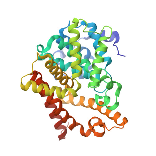
    Phosphodiesterases (PDEs) hydrolyze the second messengers cAMP and cGMP. It remains unknown how individual PDE families selectively recognize cAMP and cGMP. This work reports structural studies on substrate specificity. The crystal structures of the catalytic domains of the D674A and D564N mutants of PDE10A2 in complex with cAMP and cGMP reveal that two substrates bind to the active site with the same syn configuration but different orientations and interactions. The products AMP and GMP bind PDE10A2 with the anti configuration and interact with both divalent metals, in contrast to no direct contact of the substrates. The structures suggest that the syn configurations of cAMP and cGMP are the genuine substrates for PDE10 and the specificity is achieved through the different interactions and conformations of the substrates. The PDE10A2 structures also show that the conformation of the invariant glutamine is locked by two hydrogen bonds and is unlikely to switch for substrate recognition. Sequence alignment shows a potential pocket, in which variation of amino acids across PDE families defines the size and shape of the pocket and thus determines the substrate specificity.


  • Organizational Affiliation

    Department of Biochemistry and Biophysics and Lineberger Comprehensive Cancer Center, University of North Carolina, Chapel Hill, NC 27599-7260, USA.


Macromolecules
Find similar proteins by:  (by identity cutoff)  |  3D Structure
Entity ID: 1
MoleculeChains Sequence LengthOrganismDetailsImage
cAMP and cAMP-inhibited cGMP 3',5'-cyclic phosphodiesterase 10A
A, B
331Homo sapiensMutation(s): 1 
Gene Names: PDE10A
EC: 3.1.4.17
UniProt & NIH Common Fund Data Resources
Find proteins for Q9Y233 (Homo sapiens)
Explore Q9Y233 
Go to UniProtKB:  Q9Y233
PHAROS:  Q9Y233
GTEx:  ENSG00000112541 
Entity Groups  
Sequence Clusters30% Identity50% Identity70% Identity90% Identity95% Identity100% Identity
UniProt GroupQ9Y233
Sequence Annotations
Expand
  • Reference Sequence
Experimental Data & Validation

Experimental Data

  • Method: X-RAY DIFFRACTION
  • Resolution: 1.56 Å
  • R-Value Free: 0.228 
  • R-Value Work: 0.209 
  • Space Group: P 21 21 21
Unit Cell:
Length ( Å )Angle ( ˚ )
a = 51.441α = 90
b = 82.205β = 90
c = 155.242γ = 90
Software Package:
Software NamePurpose
ADSCdata collection
AMoREphasing
CNSrefinement
HKL-2000data reduction
HKL-2000data scaling

Structure Validation

View Full Validation Report


Entry History 

Deposition Data

Revision History  (Full details and data files)

  • Version 1.0: 2007-03-20
    Type: Initial release
  • Version 1.1: 2008-05-01
    Changes: Version format compliance
  • Version 1.2: 2011-07-13
    Changes: Version format compliance
  • Version 1.3: 2021-10-20
    Changes: Database references, Derived calculations
  • Version 1.4: 2024-02-21
    Changes: Data collection