2OU9

Structure of Spin-labeled T4 Lysozyme Mutant T115R1/R119A


Experimental Data Snapshot

  • Method: X-RAY DIFFRACTION
  • Resolution: 1.55 Å
  • R-Value Free: 0.220 
  • R-Value Work: 0.159 
  • R-Value Observed: 0.199 

wwPDB Validation   3D Report Full Report


This is version 1.6 of the entry. See complete history


Literature

Structural determinants of nitroxide motion in spin-labeled proteins: Tertiary contact and solvent-inaccessible sites in helix G of T4 lysozyme.

Guo, Z.Cascio, D.Hideg, K.Kalai, T.Hubbell, W.L.

(2007) Protein Sci 16: 1069-1086

  • DOI: https://doi.org/10.1110/ps.062739107
  • Primary Citation of Related Structures:  
    2IGC, 2NTG, 2NTH, 2OU8, 2OU9

  • PubMed Abstract: 

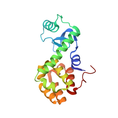
    A nitroxide side chain (R1) has been substituted at single sites along a helix-turn-helix motif in T4 lysozyme (residues 114-135). Together with previously published data, the new sites reported complete a continuous scan through the motif. Mutants with R1 at sites 115 and 118 were selected for crystallographic analysis to identify the structural origins of the corresponding two-component EPR spectra. At 115, R1 is shown to occupy two rotamers in the room temperature crystal structure, one of which has not been previously reported. The two components in the EPR spectrum apparently arise from differential interactions of the two rotamers with the surrounding structure, the most important of which is a hydrophobic interaction of the nitroxide ring. Interestingly, the crystal structure at 100 K reveals a single rotamer, emphasizing the possibility of rotamer selection in low-temperature crystal structures. Residue 118 is at a solvent-inaccessible site in the protein core, and the structure of 118R1, the first reported for the R1 side chain at a buried site, reveals how the side chain is accommodated in an overpacked core.


  • Organizational Affiliation

    Jules Stein Eye Institute and Department of Chemistry and Biochemistry, University of California, Los Angeles, California 90095-7008, USA.


Macromolecules
Find similar proteins by:  (by identity cutoff)  |  3D Structure
Entity ID: 1
MoleculeChains Sequence LengthOrganismDetailsImage
Lysozyme164Tequatrovirus T4Mutation(s): 3 
Gene Names: E
EC: 3.2.1.17
UniProt
Find proteins for P00720 (Enterobacteria phage T4)
Explore P00720 
Go to UniProtKB:  P00720
Entity Groups  
Sequence Clusters30% Identity50% Identity70% Identity90% Identity95% Identity100% Identity
UniProt GroupP00720
Sequence Annotations
Expand
  • Reference Sequence
Small Molecules
Ligands 1 Unique
IDChains Name / Formula / InChI Key2D Diagram3D Interactions
MTN
Query on MTN

Download Ideal Coordinates CCD File 
B [auth A]S-[(1-oxyl-2,2,5,5-tetramethyl-2,5-dihydro-1H-pyrrol-3-yl)methyl] methanesulfonothioate
C10 H18 N O3 S2
MXZPGYFBZHBAQM-UHFFFAOYSA-N
Experimental Data & Validation

Experimental Data

  • Method: X-RAY DIFFRACTION
  • Resolution: 1.55 Å
  • R-Value Free: 0.220 
  • R-Value Work: 0.159 
  • R-Value Observed: 0.199 
  • Space Group: P 32 2 1
Unit Cell:
Length ( Å )Angle ( ˚ )
a = 60.406α = 90
b = 60.406β = 90
c = 96.373γ = 120
Software Package:
Software NamePurpose
DENZOdata reduction
SCALEPACKdata scaling
SHELXrefinement
PDB_EXTRACTdata extraction
CrystalCleardata collection
EPMRphasing
SHELXL-97refinement

Structure Validation

View Full Validation Report


Entry History 

Deposition Data

Revision History  (Full details and data files)

  • Version 1.0: 2007-06-12
    Type: Initial release
  • Version 1.1: 2008-05-01
    Changes: Version format compliance
  • Version 1.2: 2011-07-13
    Changes: Version format compliance
  • Version 1.3: 2011-07-27
    Changes: Non-polymer description
  • Version 1.4: 2017-10-18
    Changes: Advisory, Refinement description
  • Version 1.5: 2021-10-20
    Changes: Advisory, Database references, Derived calculations
  • Version 1.6: 2023-08-30
    Changes: Data collection, Refinement description