2OU0

1-methylpyrrole in complex with T4 Lysozyme L99A


Experimental Data Snapshot

  • Method: X-RAY DIFFRACTION
  • Resolution: 1.94 Å
  • R-Value Free: 0.234 
  • R-Value Work: 0.191 
  • R-Value Observed: 0.193 

wwPDB Validation   3D Report Full Report


This is version 1.4 of the entry. See complete history


Literature

Predicting absolute ligand binding free energies to a simple model site.

Mobley, D.L.Graves, A.P.Chodera, J.D.McReynolds, A.C.Shoichet, B.K.Dill, K.A.

(2007) J Mol Biol 371: 1118-1134

  • DOI: https://doi.org/10.1016/j.jmb.2007.06.002
  • Primary Citation of Related Structures:  
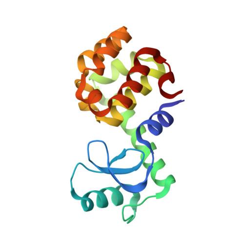
    2OTY, 2OTZ, 2OU0

  • PubMed Abstract: 

    A central challenge in structure-based ligand design is the accurate prediction of binding free energies. Here we apply alchemical free energy calculations in explicit solvent to predict ligand binding in a model cavity in T4 lysozyme. Even in this simple site, there are challenges. We made systematic improvements, beginning with single poses from docking, then including multiple poses, additional protein conformational changes, and using an improved charge model. Computed absolute binding free energies had an RMS error of 1.9 kcal/mol relative to previously determined experimental values. In blind prospective tests, the methods correctly discriminated between several true ligands and decoys in a set of putative binders identified by docking. In these prospective tests, the RMS error in predicted binding free energies relative to those subsequently determined experimentally was only 0.6 kcal/mol. X-ray crystal structures of the new ligands bound in the cavity corresponded closely to predictions from the free energy calculations, but sometimes differed from those predicted by docking. Finally, we examined the impact of holding the protein rigid, as in docking, with a view to learning how approximations made in docking affect accuracy and how they may be improved.


  • Organizational Affiliation

    Department of Pharmaceutical Chemistry, University of California at San Francisco, San Francisco, CA 94143-2518, USA.


Macromolecules
Find similar proteins by:  (by identity cutoff)  |  3D Structure
Entity ID: 1
MoleculeChains Sequence LengthOrganismDetailsImage
LysozymeA [auth X]162Tequatrovirus T4Mutation(s): 3 
Gene Names: E
EC: 3.2.1.17
UniProt
Find proteins for P00720 (Enterobacteria phage T4)
Explore P00720 
Go to UniProtKB:  P00720
Entity Groups  
Sequence Clusters30% Identity50% Identity70% Identity90% Identity95% Identity100% Identity
UniProt GroupP00720
Sequence Annotations
Expand
  • Reference Sequence
Experimental Data & Validation

Experimental Data

  • Method: X-RAY DIFFRACTION
  • Resolution: 1.94 Å
  • R-Value Free: 0.234 
  • R-Value Work: 0.191 
  • R-Value Observed: 0.193 
  • Space Group: P 32 2 1
Unit Cell:
Length ( Å )Angle ( ˚ )
a = 60.205α = 90
b = 60.205β = 90
c = 96.372γ = 120
Software Package:
Software NamePurpose
DENZOdata reduction
SCALEPACKdata scaling
REFMACrefinement
PDB_EXTRACTdata extraction
HKL-2000data collection
HKL-2000data reduction
REFMACphasing

Structure Validation

View Full Validation Report


Entry History 

Deposition Data

Revision History  (Full details and data files)

  • Version 1.0: 2007-08-07
    Type: Initial release
  • Version 1.1: 2011-07-13
    Changes: Version format compliance
  • Version 1.2: 2017-10-18
    Changes: Refinement description
  • Version 1.3: 2021-10-20
    Changes: Database references, Derived calculations
  • Version 1.4: 2023-08-30
    Changes: Data collection, Refinement description