2OMG

Structure of human insulin cocrystallized with protamine and urea


Experimental Data Snapshot

  • Method: X-RAY DIFFRACTION
  • Resolution: 1.52 Å
  • R-Value Free: 0.209 
  • R-Value Work: 0.183 
  • R-Value Observed: 0.184 

wwPDB Validation   3D Report Full Report


This is version 1.4 of the entry. See complete history


Literature

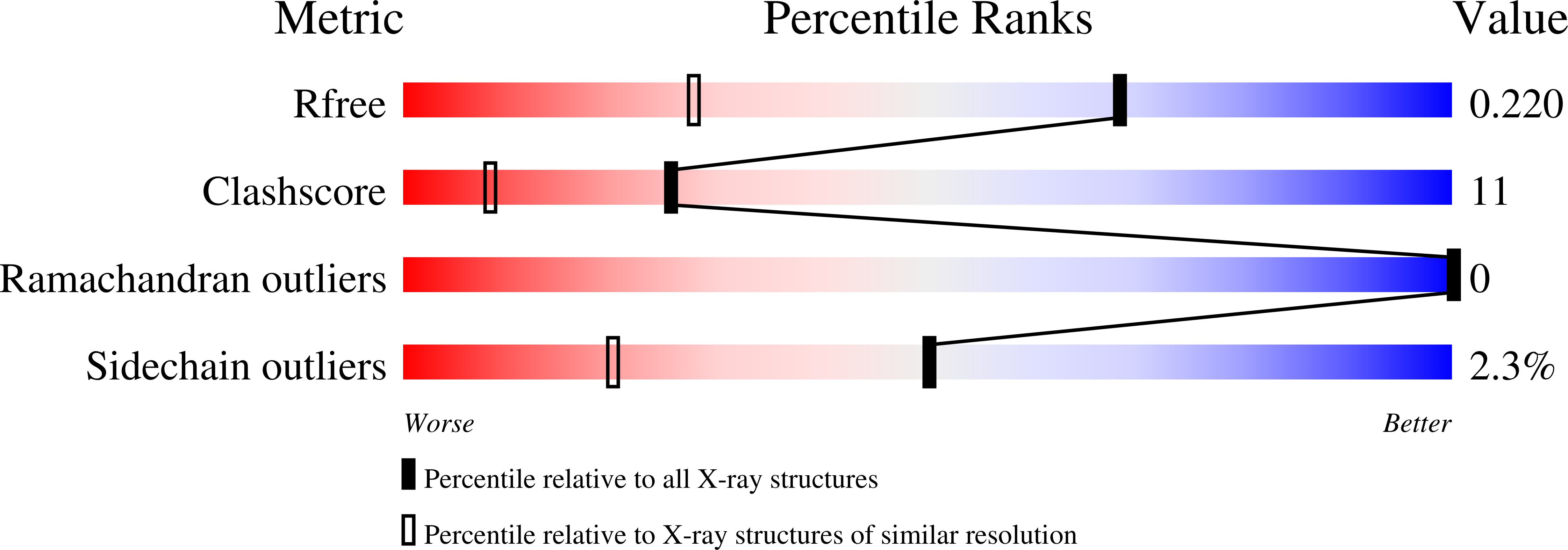
Structural characterization of insulin NPH formulations.

Norrman, M.Hubalek, F.Schluckebier, G.

(2007) Eur J Pharm Sci 30: 414-423

  • DOI: https://doi.org/10.1016/j.ejps.2007.01.003
  • Primary Citation of Related Structures:  
    2OMG, 2OMH, 2OMI

  • PubMed Abstract: 

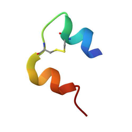
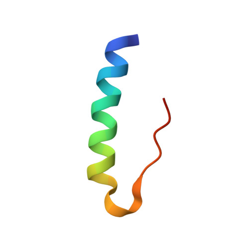
    Insulin NPH (neutral protamine hagedorn) has for long been one of the most important therapeutic formulations for the treatment of diabetes. The protracted action profile of NPH formulations is gained from crystallizing insulin with zinc in the presence of the basic poly-arginine peptide protamine. In spite of its long history and successful use, the binding mode of the insulin-protamine complex is not known. In this study, three different systems were used to study protamine binding to insulin. In the first system, crystals of an insulin-protamine complex grown in the presence of urea and diffracting to 1.5A resolution were analyzed. In the second system, a shorter peptide consisting of 12 arginine residues was co-crystallized with insulin in order to reduce the flexibility and thereby improve the electron density of the peptide. Both systems yielded data to a significantly higher resolution than obtained previously. In addition, a third system was analyzed where crystals of insulin and protamine were grown in the absence of urea, with conditions closely resembling the pharmaceutical formulation. Data from these NPH microcrystals could for the first time be collected to 2.2A resolution at a micro focused X-ray beamline. Analysis of all three crystal forms reveal potential protamine density located close to the solvent channel leading to the centrally located zinc atoms in the insulin hexamer and support that protamine binds to insulin in a not well defined conformation.


  • Organizational Affiliation

    Diabetes Protein Engineering, Novo Nordisk A/S, Novo Nordisk Park, 2760 Måløv, Denmark.


Macromolecules

Find similar proteins by:  Sequence   |   3D Structure  

Entity ID: 1
MoleculeChains Sequence LengthOrganismDetailsImage
Insulin A chain
A, C, E
21Homo sapiensMutation(s): 0 
UniProt & NIH Common Fund Data Resources
Find proteins for P01308 (Homo sapiens)
Explore P01308 
Go to UniProtKB:  P01308
PHAROS:  P01308
GTEx:  ENSG00000254647 
Entity Groups  
Sequence Clusters30% Identity50% Identity70% Identity90% Identity95% Identity100% Identity
UniProt GroupP01308
Sequence Annotations
Expand
  • Reference Sequence
Find similar proteins by:  (by identity cutoff)  |  3D Structure
Entity ID: 2
MoleculeChains Sequence LengthOrganismDetailsImage
Insulin B chain
B, D, F
30Homo sapiensMutation(s): 0 
UniProt & NIH Common Fund Data Resources
Find proteins for P01308 (Homo sapiens)
Explore P01308 
Go to UniProtKB:  P01308
PHAROS:  P01308
GTEx:  ENSG00000254647 
Entity Groups  
Sequence Clusters30% Identity50% Identity70% Identity90% Identity95% Identity100% Identity
UniProt GroupP01308
Sequence Annotations
Expand
  • Reference Sequence
Small Molecules
Ligands 6 Unique
IDChains Name / Formula / InChI Key2D Diagram3D Interactions
CRS
Query on CRS

Download Ideal Coordinates CCD File 
H [auth A],
P [auth C],
T [auth E]
M-CRESOL
C7 H8 O
RLSSMJSEOOYNOY-UHFFFAOYSA-N
ZN
Query on ZN

Download Ideal Coordinates CCD File 
L [auth B]ZINC ION
Zn
PTFCDOFLOPIGGS-UHFFFAOYSA-N
URE
Query on URE

Download Ideal Coordinates CCD File 
I [auth A]
J [auth A]
N [auth B]
Q [auth C]
R [auth D]
I [auth A],
J [auth A],
N [auth B],
Q [auth C],
R [auth D],
U [auth E]
UREA
C H4 N2 O
XSQUKJJJFZCRTK-UHFFFAOYSA-N
ARF
Query on ARF

Download Ideal Coordinates CCD File 
K [auth A],
O [auth B],
S [auth D],
V [auth E],
W [auth F]
FORMAMIDE
C H3 N O
ZHNUHDYFZUAESO-UHFFFAOYSA-N
CL
Query on CL

Download Ideal Coordinates CCD File 
M [auth B]CHLORIDE ION
Cl
VEXZGXHMUGYJMC-UHFFFAOYSA-M
NA
Query on NA

Download Ideal Coordinates CCD File 
G [auth A]SODIUM ION
Na
FKNQFGJONOIPTF-UHFFFAOYSA-N
Experimental Data & Validation

Experimental Data

  • Method: X-RAY DIFFRACTION
  • Resolution: 1.52 Å
  • R-Value Free: 0.209 
  • R-Value Work: 0.183 
  • R-Value Observed: 0.184 
  • Space Group: P 43 21 2
Unit Cell:
Length ( Å )Angle ( ˚ )
a = 61.66α = 90
b = 61.66β = 90
c = 85.54γ = 90
Software Package:
Software NamePurpose
REFMACrefinement
XDSdata reduction
XSCALEdata scaling
MOLREPphasing

Structure Validation

View Full Validation Report


Entry History 

Deposition Data

Revision History  (Full details and data files)

  • Version 1.0: 2007-03-27
    Type: Initial release
  • Version 1.1: 2008-05-01
    Changes: Version format compliance
  • Version 1.2: 2011-07-13
    Changes: Advisory, Derived calculations, Version format compliance
  • Version 1.3: 2018-03-07
    Changes: Data collection
  • Version 1.4: 2023-12-27
    Changes: Data collection, Database references, Derived calculations