2OLB

OLIGOPEPTIDE BINDING PROTEIN (OPPA) COMPLEXED WITH TRI-LYSINE


Experimental Data Snapshot

  • Method: X-RAY DIFFRACTION
  • Resolution: 1.40 Å
  • R-Value Observed: 0.183 

wwPDB Validation   3D Report Full Report


This is version 1.2 of the entry. See complete history


Literature

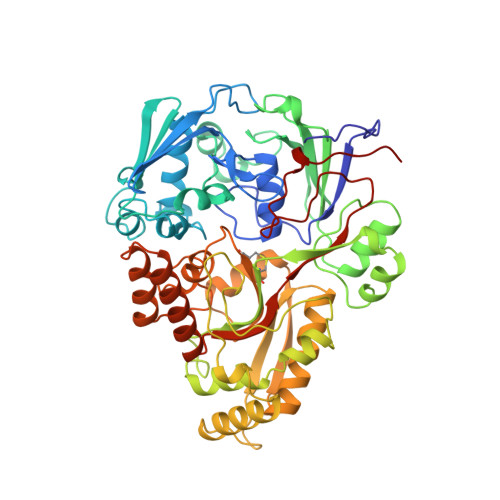
The crystal structures of the oligopeptide-binding protein OppA complexed with tripeptide and tetrapeptide ligands.

Tame, J.R.Dodson, E.J.Murshudov, G.Higgins, C.F.Wilkinson, A.J.

(1995) Structure 3: 1395-1406

  • DOI: https://doi.org/10.1016/s0969-2126(01)00276-3
  • Primary Citation of Related Structures:  
    1OLC, 2OLB

  • PubMed Abstract: 

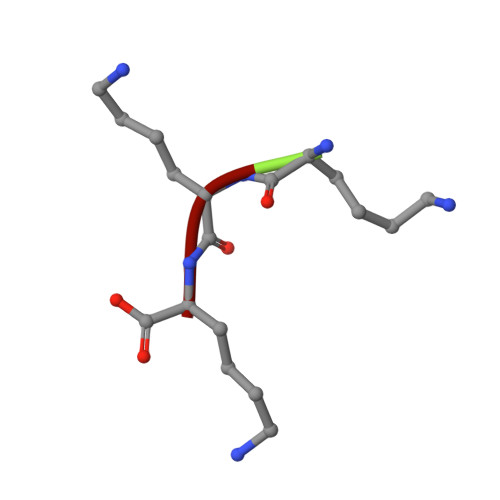
    The periplasmic oligopeptide-binding protein OppA has a remarkably broad substrate specificity, binding peptides of two or five amino-acid residues with high affinity, but little regard to sequence. It is therefore an ideal system for studying how different chemical groups can be accommodated in a protein interior. The ability of the protein to bind peptides of different lengths has been studied by co-crystallising it with different ligands. Crystals of OppA from Salmonella typhimurium complexed with the peptides Lys-Lys-Lys (KKK) and Lys-Lys-Lys-Ala (KKKA) have been grown in the presence of uranyl ions which form important crystal contacts. These structures have been refined to 1.4 A and 2.1 A, respectively. The ligands are completely enclosed, their side chains pointing into large hydrated cavities and making few strong interactions with the protein. Tight peptide binding by OppA arises from strong hydrogen bonding and electrostatic interactions between the protein and the main chain of the ligand. Different basic side chains on the protein form salt bridges with the C terminus of peptide ligands of different lengths.


  • Organizational Affiliation

    Department of Chemistry, University of York, UK.


Macromolecules
Find similar proteins by:  (by identity cutoff)  |  3D Structure
Entity ID: 1
MoleculeChains Sequence LengthOrganismDetailsImage
OLIGO-PEPTIDE BINDING PROTEIN517Salmonella enterica subsp. enterica serovar TyphimuriumMutation(s): 0 
UniProt
Find proteins for P06202 (Salmonella typhimurium (strain LT2 / SGSC1412 / ATCC 700720))
Explore P06202 
Go to UniProtKB:  P06202
Entity Groups  
Sequence Clusters30% Identity50% Identity70% Identity90% Identity95% Identity100% Identity
UniProt GroupP06202
Sequence Annotations
Expand
  • Reference Sequence

Find similar proteins by:  Sequence   |   3D Structure  

Entity ID: 2
MoleculeChains Sequence LengthOrganismDetailsImage
TRIPEPTIDE LYS-LYS-LYS3N/AMutation(s): 0 
Sequence Annotations
Expand
  • Reference Sequence
Small Molecules
Ligands 2 Unique
IDChains Name / Formula / InChI Key2D Diagram3D Interactions
IUM
Query on IUM

Download Ideal Coordinates CCD File 
C [auth A]
D [auth A]
E [auth A]
F [auth A]
G [auth A]
C [auth A],
D [auth A],
E [auth A],
F [auth A],
G [auth A],
H [auth A],
I [auth A],
J [auth A]
URANYL (VI) ION
O2 U
IZLHQJIUOZHTLW-UHFFFAOYSA-N
ACT
Query on ACT

Download Ideal Coordinates CCD File 
K [auth A]
L [auth A]
M [auth A]
N [auth A]
O [auth A]
K [auth A],
L [auth A],
M [auth A],
N [auth A],
O [auth A],
P [auth A]
ACETATE ION
C2 H3 O2
QTBSBXVTEAMEQO-UHFFFAOYSA-M
Experimental Data & Validation

Experimental Data

  • Method: X-RAY DIFFRACTION
  • Resolution: 1.40 Å
  • R-Value Observed: 0.183 
  • Space Group: P 21 21 21
Unit Cell:
Length ( Å )Angle ( ˚ )
a = 109.19α = 90
b = 76.04β = 90
c = 70.28γ = 90
Software Package:
Software NamePurpose
XDSdata scaling
PROLSQrefinement
MOSFLMdata reduction
XDSdata reduction

Structure Validation

View Full Validation Report


Entry History 

Deposition Data

Revision History  (Full details and data files)

  • Version 1.0: 1996-01-29
    Type: Initial release
  • Version 1.1: 2008-03-03
    Changes: Version format compliance
  • Version 1.2: 2011-07-13
    Changes: Version format compliance