2OJP

The crystal structure of a dimeric mutant of Dihydrodipicolinate synthase from E.coli- DHDPS-L197Y


Experimental Data Snapshot

  • Method: X-RAY DIFFRACTION
  • Resolution: 1.70 Å
  • R-Value Free: 0.228 
  • R-Value Work: 0.185 
  • R-Value Observed: 0.186 

wwPDB Validation   3D Report Full Report


This is version 1.5 of the entry. See complete history


Literature

Evolution of quaternary structure in a homotetrameric enzyme.

Griffin, M.D.Dobson, R.C.Pearce, F.G.Antonio, L.Whitten, A.E.Liew, C.K.Mackay, J.P.Trewhella, J.Jameson, G.B.Perugini, M.A.Gerrard, J.A.

(2008) J Mol Biol 380: 691-703

  • DOI: https://doi.org/10.1016/j.jmb.2008.05.038
  • Primary Citation of Related Structures:  
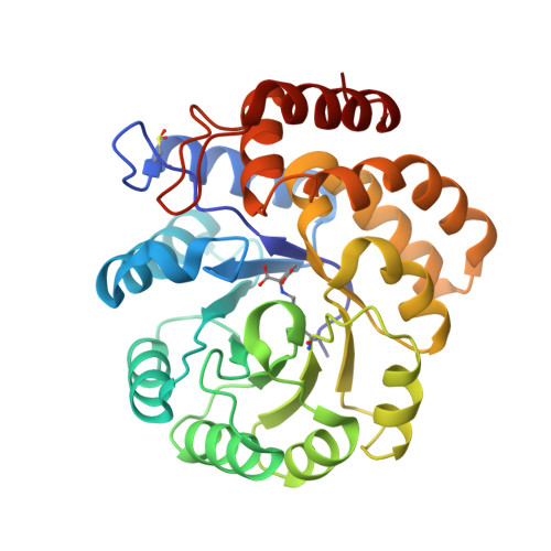
    2OJP

  • PubMed Abstract: 

    Dihydrodipicolinate synthase (DHDPS) is an essential enzyme in (S)-lysine biosynthesis and an important antibiotic target. All X-ray crystal structures solved to date reveal a homotetrameric enzyme. In order to explore the role of this quaternary structure, dimeric variants of Escherichia coli DHDPS were engineered and their properties were compared to those of the wild-type tetrameric form. X-ray crystallography reveals that the active site is not disturbed when the quaternary structure is disrupted. However, the activity of the dimeric enzymes in solution is substantially reduced, and a tetrahedral adduct of a substrate analogue is observed to be trapped at the active site in the crystal form. Remarkably, heating the dimeric enzymes increases activity. We propose that the homotetrameric structure of DHDPS reduces dynamic fluctuations present in the dimeric forms and increases specificity for the first substrate, pyruvate. By restricting motion in a key catalytic motif, a competing, non-productive reaction with a substrate analogue is avoided. Small-angle X-ray scattering and mutagenesis data, together with a B-factor analysis of the crystal structures, support this hypothesis and lead to the suggestion that in at least some cases, the evolution of quaternary enzyme structures might serve to optimise the dynamic properties of the protein subunits.


  • Organizational Affiliation

    School of Biological Sciences, University of Canterbury, Christchurch 8140, New Zealand.


Macromolecules
Find similar proteins by:  (by identity cutoff)  |  3D Structure
Entity ID: 1
MoleculeChains Sequence LengthOrganismDetailsImage
Dihydrodipicolinate synthase
A, B
292Escherichia coliMutation(s): 1 
Gene Names: dapA
EC: 4.2.1.52
UniProt
Find proteins for P0A6L2 (Escherichia coli (strain K12))
Explore P0A6L2 
Go to UniProtKB:  P0A6L2
Entity Groups  
Sequence Clusters30% Identity50% Identity70% Identity90% Identity95% Identity100% Identity
UniProt GroupP0A6L2
Sequence Annotations
Expand
  • Reference Sequence
Small Molecules
Modified Residues  2 Unique
IDChains TypeFormula2D DiagramParent
CSD
Query on CSD
A, B
L-PEPTIDE LINKINGC3 H7 N O4 SCYS
KGC
Query on KGC
A, B
L-PEPTIDE LINKINGC11 H18 N2 O6LYS
Experimental Data & Validation

Experimental Data

  • Method: X-RAY DIFFRACTION
  • Resolution: 1.70 Å
  • R-Value Free: 0.228 
  • R-Value Work: 0.185 
  • R-Value Observed: 0.186 
  • Space Group: P 21 21 21
Unit Cell:
Length ( Å )Angle ( ˚ )
a = 60.482α = 90
b = 106.705β = 90
c = 120.657γ = 90
Software Package:
Software NamePurpose
AMoREphasing
REFMACrefinement
d*TREKdata scaling

Structure Validation

View Full Validation Report


Entry History 

Deposition Data

Revision History  (Full details and data files)

  • Version 1.0: 2008-01-01
    Type: Initial release
  • Version 1.1: 2011-07-13
    Changes: Non-polymer description, Version format compliance
  • Version 1.2: 2014-11-19
    Changes: Non-polymer description
  • Version 1.3: 2017-10-18
    Changes: Refinement description
  • Version 1.4: 2021-10-20
    Changes: Database references, Derived calculations
  • Version 1.5: 2023-12-27
    Changes: Data collection