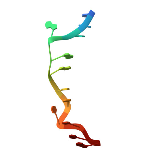
2O1I

RH(BPY)2CHRYSI complexed to mismatched DNA


Experimental Data Snapshot

  • Method: X-RAY DIFFRACTION
  • Resolution: 1.10 Å
  • R-Value Free: 0.204 

wwPDB Validation   3D Report Full Report


Ligand Structure Quality Assessment 


This is version 1.3 of the entry. See complete history


Literature

Insights into finding a mismatch through the structure of a mispaired DNA bound by a rhodium intercalator.

Pierre, V.C.Kaiser, J.T.Barton, J.K.

(2007) Proc Natl Acad Sci U S A 104: 429-434

  • DOI: https://doi.org/10.1073/pnas.0610170104
  • Primary Citation of Related Structures:  
    2O1I

  • PubMed Abstract: 

    We report the 1.1-A resolution crystal structure of a bulky rhodium complex bound to two different DNA sites, mismatched and matched in the oligonucleotide 5'-(dCGGAAATTCCCG)2-3'. At the AC mismatch site, the structure reveals ligand insertion from the minor groove with ejection of both mismatched bases and elucidates how destabilized mispairs in DNA may be recognized. This unique binding mode contrasts with major groove intercalation, observed at a matched site, where doubling of the base pair rise accommodates stacking of the intercalator. Mass spectral analysis reveals different photocleavage products associated with the two binding modes in the crystal, with only products characteristic of mismatch binding in solution. This structure, illustrating two clearly distinct binding modes for a molecule with DNA, provides a rationale for the interrogation and detection of mismatches.


  • Organizational Affiliation

    Division of Chemistry and Chemical Engineering, California Institute of Technology, Pasadena, CA 91125, USA.


Macromolecules

Find similar nucleic acids by:  Sequence   |   3D Structure  

Entity ID: 1
MoleculeChains LengthOrganismImage
5'-D(*CP*GP*GP*AP*AP*AP*TP*TP*CP*CP*CP*G)-3'12N/A
Sequence Annotations
Expand
  • Reference Sequence
Small Molecules
Ligands 1 Unique
IDChains Name / Formula / InChI Key2D Diagram3D Interactions
R1C
Query on R1C

Download Ideal Coordinates CCD File 
B [auth A],
C [auth A],
D [auth A]
bis(2,2'-bipyridine-kappa~2~N~1~,N~1'~)[chrysene-5,6-diiminato(2-)-kappa~2~N,N']rhodium(4+)
C38 H26 N6 Rh
UKDZOHAQCIISRY-UHFFFAOYSA-N
Experimental Data & Validation

Experimental Data

  • Method: X-RAY DIFFRACTION
  • Resolution: 1.10 Å
  • R-Value Free: 0.204 
  • Space Group: P 43 21 2
Unit Cell:
Length ( Å )Angle ( ˚ )
a = 38.74α = 90
b = 38.74β = 90
c = 57.56γ = 90
Software Package:
Software NamePurpose
SHELXmodel building
SHELXL-97refinement
Blu-Icedata collection
MOSFLMdata reduction
CCP4data scaling
SHELXDphasing

Structure Validation

View Full Validation Report


Ligand Structure Quality Assessment 


Entry History 

Deposition Data

Revision History  (Full details and data files)

  • Version 1.0: 2007-01-09
    Type: Initial release
  • Version 1.1: 2008-05-01
    Changes: Version format compliance
  • Version 1.2: 2011-07-13
    Changes: Version format compliance
  • Version 1.3: 2023-12-27
    Changes: Data collection, Database references, Derived calculations