2NSY

CRYSTAL STRUCTURE OF NH3-DEPENDENT NAD+ SYNTHETASE FROM BACILLUS SUBTILIS IN COMPLEX WITH NAD-ADENYLATE


Experimental Data Snapshot

  • Method: X-RAY DIFFRACTION
  • Resolution: 2.00 Å
  • R-Value Free: 0.180 
  • R-Value Work: 0.155 

wwPDB Validation   3D Report Full Report


This is version 1.4 of the entry. See complete history


Literature

A novel deamido-NAD+-binding site revealed by the trapped NAD-adenylate intermediate in the NAD+ synthetase structure.

Rizzi, M.Bolognesi, M.Coda, A.

(1998) Structure 6: 1129-1140

  • DOI: https://doi.org/10.1016/s0969-2126(98)00114-2
  • Primary Citation of Related Structures:  
    2NSY

  • PubMed Abstract: 

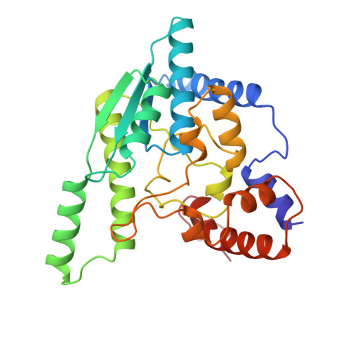
    Nicotinamide adenine dinucleotide (NAD+) has a central role in life processes. The ubiquitous enzyme NAD+ synthetase catalyzes a key step in NAD+ biosynthesis, transforming deamido-NAD+ into NAD+ by a two-step reaction. NAD+ synthetase belongs to the amidotransferase family and has been recognized as a member of the family of N-type ATP pyrophosphatases. In order to investigate the mechanism of the reaction carried out by NAD+ synthetase we have determined a high-resolution three-dimensional structure of the Bacillus subtilis homodimeric NAD+ synthetase in complex with the trapped reaction intermediate NAD-adenylate. Two NAD-adenylate molecules and two pyrophosphate (PPi) molecules are observed in the 1.3 A resolution structure of the NAD+ synthetase-NAD-adenylate complex. Structural studies on the NAD+ synthetase-NAD-adenylate adduct and on the cation-binding sites reveal a new deamido-NAD+-binding site located at the subunit interface, locate a binuclear magnesium cluster at the ATP-binding site and, identify two monovalent cation sites, one of which may represent an ammonium-binding site. Our results suggest that two different catalytic strategies have been adopted by NAD+ synthetase in the two different steps of the reaction. During the adenylation step, no protein residues seem to be located properly to directly participate in catalysis, which is likely to be carried out with the fundamental assistance of an electron-withdrawing trimetallic constellation present in the active site. A different behavior is observed for the second step, in which an ammonium ion is the binding species. In this step, Asp173 is a key residue in both deprotonation of the primarily bound ammonium ion, and stabilization of the tetrahedral transition-state intermediate. Moreover, the structural data suggest that product release can take place only after all substrates are bound to the enzyme, and product release is ultimately controlled by the conformation adopted by two mobile loops.


  • Organizational Affiliation

    Department of Pharmaceutical Science and Technology University of Torino, Italy. rizzi@ipvgen.unipv.it


Macromolecules
Find similar proteins by:  (by identity cutoff)  |  3D Structure
Entity ID: 1
MoleculeChains Sequence LengthOrganismDetailsImage
PROTEIN (NAD SYNTHETASE)
A, B
271Bacillus subtilisMutation(s): 0 
EC: 6.3.5.1
UniProt
Find proteins for P08164 (Bacillus subtilis (strain 168))
Explore P08164 
Go to UniProtKB:  P08164
Entity Groups  
Sequence Clusters30% Identity50% Identity70% Identity90% Identity95% Identity100% Identity
UniProt GroupP08164
Sequence Annotations
Expand
  • Reference Sequence
Small Molecules
Ligands 5 Unique
IDChains Name / Formula / InChI Key2D Diagram3D Interactions
NAD
Query on NAD

Download Ideal Coordinates CCD File 
F [auth A],
L [auth B]
NICOTINAMIDE-ADENINE-DINUCLEOTIDE
C21 H27 N7 O14 P2
BAWFJGJZGIEFAR-NNYOXOHSSA-N
AMP
Query on AMP

Download Ideal Coordinates CCD File 
E [auth A],
K [auth B]
ADENOSINE MONOPHOSPHATE
C10 H14 N5 O7 P
UDMBCSSLTHHNCD-KQYNXXCUSA-N
POP
Query on POP

Download Ideal Coordinates CCD File 
G [auth A],
M [auth B]
PYROPHOSPHATE 2-
H2 O7 P2
XPPKVPWEQAFLFU-UHFFFAOYSA-L
GOL
Query on GOL

Download Ideal Coordinates CCD File 
H [auth A],
N [auth B]
GLYCEROL
C3 H8 O3
PEDCQBHIVMGVHV-UHFFFAOYSA-N
MG
Query on MG

Download Ideal Coordinates CCD File 
C [auth A],
D [auth A],
I [auth B],
J [auth B]
MAGNESIUM ION
Mg
JLVVSXFLKOJNIY-UHFFFAOYSA-N
Experimental Data & Validation

Experimental Data

  • Method: X-RAY DIFFRACTION
  • Resolution: 2.00 Å
  • R-Value Free: 0.180 
  • R-Value Work: 0.155 
  • Space Group: P 1 21 1
Unit Cell:
Length ( Å )Angle ( ˚ )
a = 52.48α = 90
b = 85.62β = 110.76
c = 60.12γ = 90
Software Package:
Software NamePurpose
CCP4model building
REFMACrefinement
MOSFLMdata reduction
CCP4data scaling
ROTAVATAdata scaling
CCP4phasing

Structure Validation

View Full Validation Report


Entry History 

Deposition Data

Revision History  (Full details and data files)

  • Version 1.0: 1999-01-13
    Type: Initial release
  • Version 1.1: 2008-04-27
    Changes: Version format compliance
  • Version 1.2: 2011-07-13
    Changes: Non-polymer description, Version format compliance
  • Version 1.3: 2017-10-04
    Changes: Refinement description
  • Version 1.4: 2023-08-30
    Changes: Data collection, Database references, Derived calculations, Refinement description