2NQC

Crystal structure of ig-like domain 23 from human filamin C


Experimental Data Snapshot

  • Method: X-RAY DIFFRACTION
  • Resolution: 2.05 Å
  • R-Value Free: 0.237 
  • R-Value Work: 0.188 
  • R-Value Observed: 0.190 

wwPDB Validation   3D Report Full Report


This is version 1.3 of the entry. See complete history


Literature

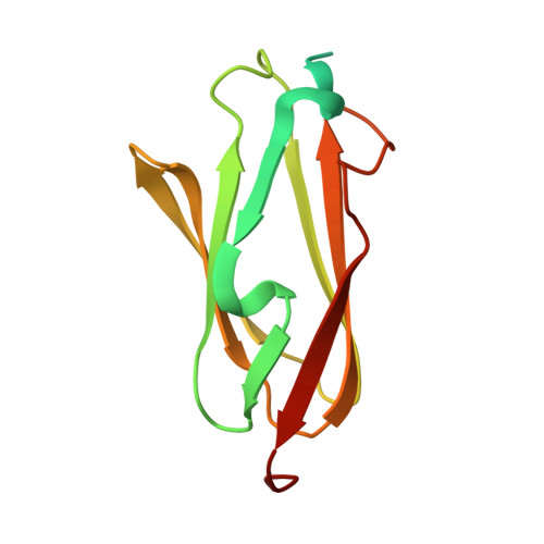
Crystal structure of human filamin C domain 23 and small angle scattering model for filamin C 23-24 dimer

Sjekloca, L.Pudas, R.Sjoeblom, B.Konarev, P.Carugo, O.Rybin, V.Kiema, T.R.Svergun, D.Ylanne, J.Djinovic-Carugo, K.

(2007) J Mol Biol 368: 1011-1023

  • DOI: https://doi.org/10.1016/j.jmb.2007.02.018
  • Primary Citation of Related Structures:  
    2NQC

  • PubMed Abstract: 

    Filamin C is a dimeric, actin-binding protein involved in organization of cortical cytoskeleton and of the sarcomere. We performed crystallographic, small-angle X-ray scattering and analytical ultracentrifugation experiments on the constructs containing carboxy-terminal domains of the protein (domains 23-24 and 19-21). The crystal structure of domain 23 of filamin C showed that the protein adopts the expected immunoglobulin (Ig)-like fold. Small-angle X-ray scattering experiments performed on filamin C tandem Ig-like domains 23 and 24 reveal a dimer that is formed by domain 24 and that domain 23 has little interactions with itself or with domain 24, while the analytical ultracentrifugation experiments showed that the filamin C domains 19-21 form elongated monomers in diluted solutions.


  • Organizational Affiliation

    International School for Advanced Studies, 34000 Trieste, Italy.


Macromolecules
Find similar proteins by:  (by identity cutoff)  |  3D Structure
Entity ID: 1
MoleculeChains Sequence LengthOrganismDetailsImage
Filamin-C138Homo sapiensMutation(s): 0 
UniProt & NIH Common Fund Data Resources
Find proteins for Q14315 (Homo sapiens)
Explore Q14315 
Go to UniProtKB:  Q14315
PHAROS:  Q14315
GTEx:  ENSG00000128591 
Entity Groups  
Sequence Clusters30% Identity50% Identity70% Identity90% Identity95% Identity100% Identity
UniProt GroupQ14315
Sequence Annotations
Expand
  • Reference Sequence
Experimental Data & Validation

Experimental Data

  • Method: X-RAY DIFFRACTION
  • Resolution: 2.05 Å
  • R-Value Free: 0.237 
  • R-Value Work: 0.188 
  • R-Value Observed: 0.190 
  • Space Group: P 31 2 1
Unit Cell:
Length ( Å )Angle ( ˚ )
a = 52.141α = 90
b = 52.141β = 90
c = 69.216γ = 120
Software Package:
Software NamePurpose
REFMACrefinement
PDB_EXTRACTdata extraction
MAR345data collection
DENZOdata reduction
SCALAdata scaling
SHARPphasing

Structure Validation

View Full Validation Report


Entry History 

Deposition Data

Revision History  (Full details and data files)

  • Version 1.0: 2007-09-11
    Type: Initial release
  • Version 1.1: 2011-07-13
    Changes: Non-polymer description, Version format compliance
  • Version 1.2: 2017-10-18
    Changes: Advisory, Refinement description
  • Version 1.3: 2023-12-27
    Changes: Advisory, Data collection, Database references, Derived calculations