2N1O

PIN1 WW domain in complex with a phosphorylated CPEB1 derived peptide


Experimental Data Snapshot

  • Method: SOLUTION NMR
  • Conformers Calculated: 120 
  • Conformers Submitted: 19 
  • Selection Criteria: structures with the lowest energy 

wwPDB Validation   3D Report Full Report


This is version 1.0 of the entry. See complete history


Literature

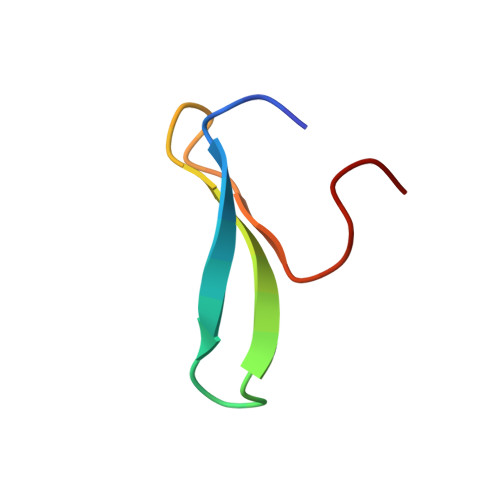
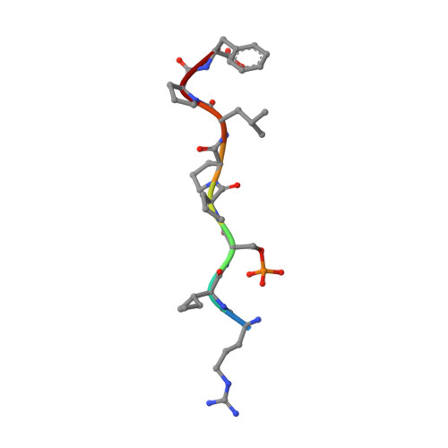
Structural Analysis of the Pin1-CPEB1 interaction and its potential role in CPEB1 degradation.

Schelhorn, C.Martin-Malpartida, P.Sunol, D.Macias, M.J.

(2015) Sci Rep 5: 14990-14990

  • DOI: https://doi.org/10.1038/srep14990
  • Primary Citation of Related Structures:  
    2N1O

  • PubMed Abstract: 

    The Cytoplasmic Polyadenylation Element Binding proteins are RNA binding proteins involved in the translational regulation of mRNA. During cell cycle progression, CPEB1 is labeled for degradation by phosphorylation-dependent ubiquitination by the SCF(β-TrCP) ligase. The peptidyl-prolyl isomerase Pin1 plays a key role in CPEB1 degradation. Conditioned by the cell cycle stage, CPEB1 and Pin1 interactions occur in a phosphorylation-independent or -dependent manner. CPEB1 contains six potential phosphorylatable Pin1 binding sites. Using a set of biophysical techniques, we discovered that the pS210 site is unique, since it displays binding activity not only to the WW domain but also to the prolyl-isomerase domain of Pin1. The NMR structure of the Pin1 WW-CPEB1 pS210 (PDB ID: 2n1o) reveals that the pSerPro motif is bound in trans configuration through contacts with amino acids located in the first turn of the WW domain and the conserved tryptophan in the β3-strand. NMR relaxation analyses of Pin1 suggest that inter-domain flexibility is conferred by the modulation of the interaction with peptides containing the pS210 site, which is essential for degradation.


  • Organizational Affiliation

    Institute for Research in Biomedicine (IRB Barcelona), The Barcelona Institute of Science and Technology (BIST), Baldiri Reixac 10, Barcelona, 08028, Spain.


Macromolecules
Find similar proteins by:  (by identity cutoff)  |  3D Structure
Entity ID: 1
MoleculeChains Sequence LengthOrganismDetailsImage
Peptidyl-prolyl cis-trans isomerase NIMA-interacting 133Homo sapiensMutation(s): 0 
Gene Names: PIN1
EC: 5.2.1.8
UniProt & NIH Common Fund Data Resources
Find proteins for Q13526 (Homo sapiens)
Explore Q13526 
Go to UniProtKB:  Q13526
PHAROS:  Q13526
GTEx:  ENSG00000127445 
Entity Groups  
Sequence Clusters30% Identity50% Identity70% Identity90% Identity95% Identity100% Identity
UniProt GroupQ13526
Sequence Annotations
Expand
  • Reference Sequence

Find similar proteins by:  Sequence   |   3D Structure  

Entity ID: 2
MoleculeChains Sequence LengthOrganismDetailsImage
Cytoplasmic polyadenylation element-binding protein 18Homo sapiensMutation(s): 0 
UniProt & NIH Common Fund Data Resources
Find proteins for Q9BZB8 (Homo sapiens)
Explore Q9BZB8 
Go to UniProtKB:  Q9BZB8
PHAROS:  Q9BZB8
GTEx:  ENSG00000214575 
Entity Groups  
UniProt GroupQ9BZB8
Sequence Annotations
Expand
  • Reference Sequence
Small Molecules
Modified Residues  1 Unique
IDChains TypeFormula2D DiagramParent
SEP
Query on SEP
B
L-PEPTIDE LINKINGC3 H8 N O6 PSER
Experimental Data & Validation

Experimental Data

  • Method: SOLUTION NMR
  • Conformers Calculated: 120 
  • Conformers Submitted: 19 
  • Selection Criteria: structures with the lowest energy 

Structure Validation

View Full Validation Report


Entry History 

Deposition Data

Revision History  (Full details and data files)

  • Version 1.0: 2015-10-28
    Type: Initial release