2MW2

Hha-H-NS46 charge zipper complex


Experimental Data Snapshot

  • Method: SOLUTION NMR
  • Conformers Calculated: 400 
  • Conformers Submitted: 10 
  • Selection Criteria: target function 

wwPDB Validation   3D Report Full Report


This is version 1.3 of the entry. See complete history


Literature

A Three-protein Charge Zipper Stabilizes a Complex Modulating Bacterial Gene Silencing.

Cordeiro, T.N.Garcia, J.Bernado, P.Millet, O.Pons, M.

(2015) J Biol Chem 290: 21200-21212

  • DOI: https://doi.org/10.1074/jbc.M114.630400
  • Primary Citation of Related Structures:  
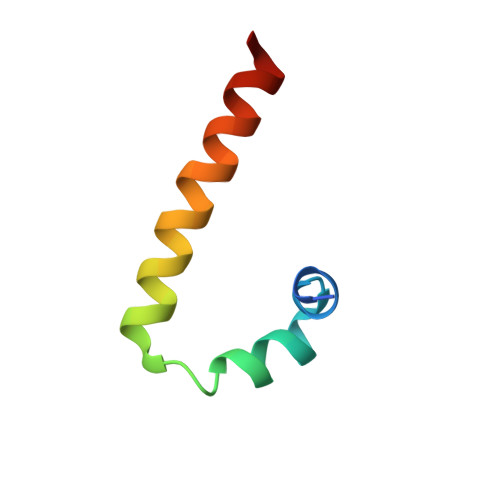
    2MW2

  • PubMed Abstract: 

    The Hha/YmoA nucleoid-associated proteins help selectively silence horizontally acquired genetic material, including pathogenicity and antibiotic resistance genes and their maintenance in the absence of selective pressure. Members of the Hha family contribute to gene silencing by binding to the N-terminal dimerization domain of H-NS and modifying its selectivity. Hha-like proteins and the H-NS N-terminal domain are unusually rich in charged residues, and their interaction is mostly electrostatic-driven but, nonetheless, highly selective. The NMR-based structural model of the complex between Hha/YmoA and the H-NS N-terminal dimerization domain reveals that the origin of the selectivity is the formation of a three-protein charge zipper with interdigitated complementary charged residues from Hha and the two units of the H-NS dimer. The free form of YmoA shows collective microsecond-millisecond dynamics that can by measured by NMR relaxation dispersion experiments and shows a linear dependence with the salt concentration. The number of residues sensing the collective dynamics and the population of the minor form increased in the presence of H-NS. Additionally, a single residue mutation in YmoA (D43N) abolished H-NS binding and the dynamics of the apo-form, suggesting the dynamics and binding are functionally related.


  • Organizational Affiliation

    From the Biomolecular NMR Laboratory, Department of Organic Chemistry, University of Barcelona, 08028 Barcelona, Spain, Centre de Biochimie Structurale, INSERM U1054, CNRS UMR 5048, Université Montpellier 1 and 2, 34092 Montpellier, France.


Macromolecules
Find similar proteins by:  (by identity cutoff)  |  3D Structure
Entity ID: 1
MoleculeChains Sequence LengthOrganismDetailsImage
Hemolysin expression-modulating protein Hha72Escherichia coli K-12Mutation(s): 0 
Gene Names: hhab0460JW0449
UniProt
Find proteins for P0ACE3 (Escherichia coli (strain K12))
Explore P0ACE3 
Go to UniProtKB:  P0ACE3
Entity Groups  
Sequence Clusters30% Identity50% Identity70% Identity90% Identity95% Identity100% Identity
UniProt GroupP0ACE3
Sequence Annotations
Expand
  • Reference Sequence
Find similar proteins by:  (by identity cutoff)  |  3D Structure
Entity ID: 2
MoleculeChains Sequence LengthOrganismDetailsImage
DNA-binding protein H-NS
B, C
47Escherichia coli K-12Mutation(s): 0 
Gene Names: 
hnsbglYcurdrdXhnsAmsyAosmZpilGtopSb1237...
hnsbglYcurdrdXhnsAmsyAosmZpilGtopSb1237JW1225

UniProt
Find proteins for P0ACF8 (Escherichia coli (strain K12))
Explore P0ACF8 
Go to UniProtKB:  P0ACF8
Entity Groups  
Sequence Clusters30% Identity50% Identity70% Identity90% Identity95% Identity100% Identity
UniProt GroupP0ACF8
Sequence Annotations
Expand
  • Reference Sequence
Experimental Data & Validation

Experimental Data

  • Method: SOLUTION NMR
  • Conformers Calculated: 400 
  • Conformers Submitted: 10 
  • Selection Criteria: target function 

Structure Validation

View Full Validation Report


Entry History 

Deposition Data

Revision History  (Full details and data files)

  • Version 1.0: 2015-07-29
    Type: Initial release
  • Version 1.1: 2016-04-27
    Changes: Structure summary
  • Version 1.2: 2018-01-10
    Changes: Database references
  • Version 1.3: 2024-05-01
    Changes: Data collection, Database references