2JB5

Fab fragment in complex with small molecule hapten, crystal form-1


Experimental Data Snapshot

  • Method: X-RAY DIFFRACTION
  • Resolution: 2.80 Å
  • R-Value Free: 0.272 
  • R-Value Work: 0.211 

wwPDB Validation   3D Report Full Report


Ligand Structure Quality Assessment 


This is version 1.3 of the entry. See complete history


Literature

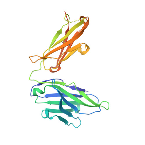
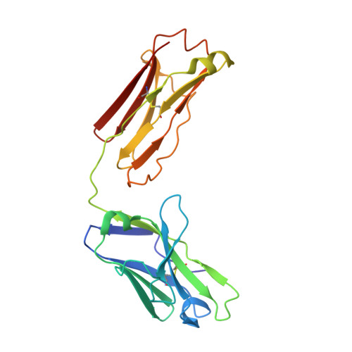
Fab Mor03268 Triggers Absorption Shift of a Diagnostic Dye Via Packaging in a Solvent-Shielded Fab Dimer Interface

Hillig, R.C.Urlinger, S.Fanghanel, J.Brocks, B.Haenel, C.Stark, Y.Sulzle, D.Svergun, D.I.Baesler, S.Malawski, G.Moosmayer, D.Menrad, A.Schirner, M.Licha, K.

(2008) J Mol Biol 377: 206

  • DOI: https://doi.org/10.1016/j.jmb.2007.12.071
  • Primary Citation of Related Structures:  
    2JB5, 2JB6

  • PubMed Abstract: 

    Molecular interactions between near-IR fluorescent probes and specific antibodies may be exploited to generate novel smart probes for diagnostic imaging. Using a new phage display technology, we developed such antibody Fab fragments with subnanomolar binding affinity for tetrasulfocyanine, a near-IR in vivo imaging agent. Unexpectedly, some Fabs induced redshifts of the dye absorption peak of up to 44 nm. This is the largest shift reported for a biological system so far. Crystal structure determination and absorption spectroscopy in the crystal in combination with microcalorimetry and small-angle X-ray scattering in solution revealed that the redshift is triggered by formation of a Fab dimer, with tetrasulfocyanine being buried in a fully closed protein cavity within the dimer interface. The derived principle of shifting the absorption peak of a symmetric dye via packaging within a Fab dimer interface may be transferred to other diagnostic fluorophores, opening the way towards smart imaging probes that change their wavelength upon interaction with an antibody.


  • Organizational Affiliation

    Bayer Schering Pharma AG, Global Drug Discovery, 13342 Berlin, Germany. roman.hillig@bayerhealthcare.com


Macromolecules
Find similar proteins by:  (by identity cutoff)  |  3D Structure
Entity ID: 1
MoleculeChains Sequence LengthOrganismDetailsImage
FAB FRAGMENT MOR03268 HEAVY CHAINA [auth H]244Homo sapiensMutation(s): 0 
Entity Groups  
Sequence Clusters30% Identity50% Identity70% Identity90% Identity95% Identity100% Identity
Sequence Annotations
Expand
  • Reference Sequence
Find similar proteins by:  (by identity cutoff)  |  3D Structure
Entity ID: 2
MoleculeChains Sequence LengthOrganismDetailsImage
FAB FRAGMENT MOR03268 LIGHT CHAINB [auth L]216Homo sapiensMutation(s): 0 
UniProt & NIH Common Fund Data Resources
Find proteins for P0DOY2 (Homo sapiens)
Explore P0DOY2 
Go to UniProtKB:  P0DOY2
GTEx:  ENSG00000211677 
Entity Groups  
Sequence Clusters30% Identity50% Identity70% Identity90% Identity95% Identity100% Identity
UniProt GroupP0DOY2
Sequence Annotations
Expand
  • Reference Sequence
Small Molecules
Ligands 1 Unique
IDChains Name / Formula / InChI Key2D Diagram3D Interactions
T5C
Query on T5C

Download Ideal Coordinates CCD File 
C [auth H]2-{(1E,3Z,5E,7E)-7-[3,3-DIMETHYL-5-SULFO-1-(2-SULFOETHYL)-1,3-DIHYDRO-2H-INDOL-2-YLIDENE]-4-METHYLHEPTA-1,3,5-TRIEN-1-YL}-3,3-DIMETHYL-5-SULFO-1-(2-SULFOETHYL)-3H-INDOLIUM
C32 H39 N2 O12 S4
WKWHOLCUESSZIQ-UHFFFAOYSA-O
Binding Affinity Annotations 
IDSourceBinding Affinity
TSC PDBBind:  2JB5 Kd: 0.6 (nM) from 1 assay(s)
Experimental Data & Validation

Experimental Data

  • Method: X-RAY DIFFRACTION
  • Resolution: 2.80 Å
  • R-Value Free: 0.272 
  • R-Value Work: 0.211 
  • Space Group: I 2 2 2
Unit Cell:
Length ( Å )Angle ( ˚ )
a = 71.949α = 90
b = 98.48β = 90
c = 153.782γ = 90
Software Package:
Software NamePurpose
CNXrefinement
HKL-2000data reduction
HKL-2000data scaling
MOLREPphasing

Structure Validation

View Full Validation Report


Ligand Structure Quality Assessment 


Entry History 

Deposition Data

Revision History  (Full details and data files)

  • Version 1.0: 2008-01-08
    Type: Initial release
  • Version 1.1: 2011-05-08
    Changes: Version format compliance
  • Version 1.2: 2011-07-13
    Changes: Version format compliance
  • Version 1.3: 2023-12-13
    Changes: Data collection, Database references, Other, Refinement description