2J44

Alpha-glucan binding by a streptococcal virulence factor


Experimental Data Snapshot

  • Method: X-RAY DIFFRACTION
  • Resolution: 2.10 Å
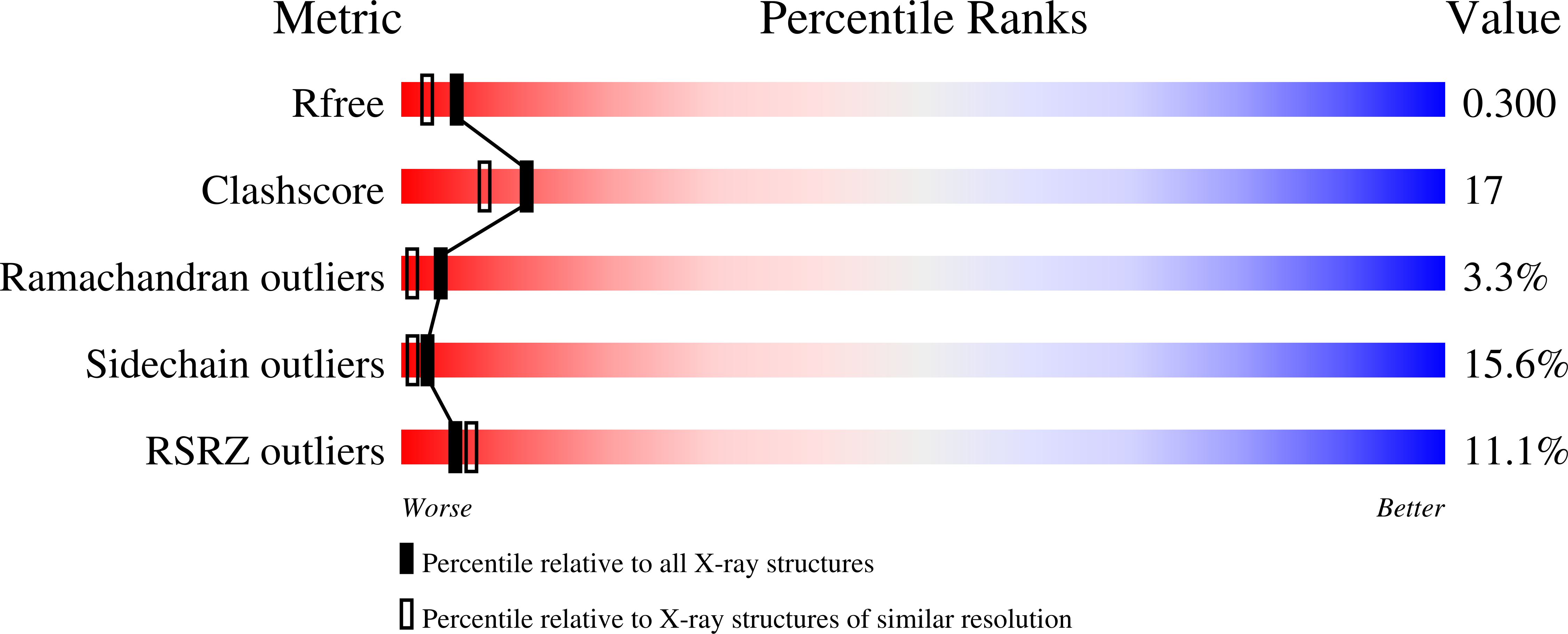
  • R-Value Free: 0.304 
  • R-Value Work: 0.221 
  • R-Value Observed: 0.225 

wwPDB Validation   3D Report Full Report


This is version 2.1 of the entry. See complete history


Literature

Identification and Structural Basis of Binding to Host Lung Glycogen by Streptococcal Virulence Factors.

Lammerts Van Bueren, A.Higgins, M.Wang, D.Burke, R.D.Boraston, A.B.

(2007) Nat Struct Mol Biol 14: 76

  • DOI: https://doi.org/10.1038/nsmb1187
  • Primary Citation of Related Structures:  
    2J43, 2J44

  • PubMed Abstract: 

    The ability of pathogenic bacteria to recognize host glycans is often essential to their virulence. Here we report structure-function studies of previously uncharacterized glycogen-binding modules in the surface-anchored pullulanases from Streptococcus pneumoniae (SpuA) and Streptococcus pyogenes (PulA). Multivalent binding to glycogen leads to a strong interaction with alveolar type II cells in mouse lung tissue. X-ray crystal structures of the binding modules reveal a novel fusion of tandem modules into single, bivalent functional domains. In addition to indicating a structural basis for multivalent attachment, the structure of the SpuA modules in complex with carbohydrate provides insight into the molecular basis for glycogen specificity. This report provides the first evidence that intracellular lung glycogen may be a novel target of pathogenic streptococci and thus provides a rationale for the identification of the streptococcal alpha-glucan-metabolizing machinery as virulence factors.


  • Organizational Affiliation

    Biochemistry & Microbiology, University of Victoria, PO Box 3055 STN CSC, Victoria, British Columbia, V8W 3P6, Canada.


Macromolecules
Find similar proteins by:  (by identity cutoff)  |  3D Structure
Entity ID: 1
MoleculeChains Sequence LengthOrganismDetailsImage
ALKALINE AMYLOPULLULANASE217Streptococcus pneumoniaeMutation(s): 0 
UniProt
Find proteins for A0A0H2UNG0 (Streptococcus pneumoniae serotype 4 (strain ATCC BAA-334 / TIGR4))
Explore A0A0H2UNG0 
Go to UniProtKB:  A0A0H2UNG0
Entity Groups  
Sequence Clusters30% Identity50% Identity70% Identity90% Identity95% Identity100% Identity
UniProt GroupA0A0H2UNG0
Sequence Annotations
Expand
  • Reference Sequence
Oligosaccharides

Help

Entity ID: 2
MoleculeChains Length2D Diagram Glycosylation3D Interactions
alpha-D-glucopyranose-(1-4)-alpha-D-glucopyranose-(1-4)-alpha-D-glucopyranose
B
3N/A
Glycosylation Resources
GlyTouCan:  G96370VA
GlyCosmos:  G96370VA
GlyGen:  G96370VA
Entity ID: 3
MoleculeChains Length2D Diagram Glycosylation3D Interactions
alpha-D-glucopyranose-(1-4)-alpha-D-glucopyranose-(1-4)-alpha-D-glucopyranose-(1-4)-alpha-D-glucopyranose
C
4N/A
Glycosylation Resources
GlyTouCan:  G87171PZ
GlyCosmos:  G87171PZ
GlyGen:  G87171PZ
Biologically Interesting Molecules (External Reference) 2 Unique
Experimental Data & Validation

Experimental Data

  • Method: X-RAY DIFFRACTION
  • Resolution: 2.10 Å
  • R-Value Free: 0.304 
  • R-Value Work: 0.221 
  • R-Value Observed: 0.225 
  • Space Group: P 21 21 2
Unit Cell:
Length ( Å )Angle ( ˚ )
a = 50.555α = 90
b = 89.124β = 90
c = 59.997γ = 90
Software Package:
Software NamePurpose
REFMACrefinement
SPYDphasing

Structure Validation

View Full Validation Report


Entry History 

Deposition Data

Revision History  (Full details and data files)

  • Version 1.0: 2006-12-11
    Type: Initial release
  • Version 1.1: 2011-05-08
    Changes: Version format compliance
  • Version 1.2: 2011-07-13
    Changes: Version format compliance
  • Version 2.0: 2020-07-29
    Type: Remediation
    Reason: Carbohydrate remediation
    Changes: Atomic model, Data collection, Derived calculations, Other, Structure summary
  • Version 2.1: 2023-12-13
    Changes: Data collection, Database references, Refinement description, Structure summary