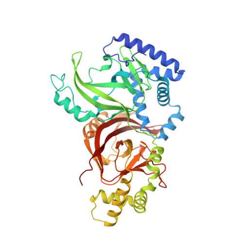
2J3X

Crystal structure of the enzymatic component C2-I of the C2-toxin from Clostridium botulinum at pH 3.0 (mut-S361R)


Experimental Data Snapshot

  • Method: X-RAY DIFFRACTION
  • Resolution: 1.75 Å
  • R-Value Free: 0.197 
  • R-Value Work: 0.168 
  • R-Value Observed: 0.169 

wwPDB Validation   3D Report Full Report


This is version 1.4 of the entry. See complete history


Literature

Structure and Action of the Binary C2 Toxin from Clostridium Botulinum.

Schleberger, C.Hochmann, H.Barth, H.Aktories, K.Schulz, G.E.

(2006) J Mol Biol 364: 705

  • DOI: https://doi.org/10.1016/j.jmb.2006.09.002
  • Primary Citation of Related Structures:  
    2J3V, 2J3X, 2J3Z, 2J42

  • PubMed Abstract: 

    C2 toxin from Clostridium botulinum is composed of the enzyme component C2-I, which ADP-ribosylates actin, and the binding and translocation component C2-II, responsible for the interaction with eukaryotic cell receptors and the following endocytosis. Three C2-I crystal structures at resolutions of up to 1.75 A are presented together with a crystal structure of C2-II at an appreciably lower resolution and a model of the prepore formed by fragment C2-IIa. The C2-I structure was determined at pH 3.0 and at pH 6.1. The structural differences are small, indicating that C2-I does not unfold, even at a pH value as low as 3.0. The ADP-ribosyl transferase activity of C2-I was determined for alpha and beta/gamma-actin and related to that of Iota toxin and of mutant S361R of C2-I that introduced the arginine observed in Iota toxin. The substantial activity differences between alpha and beta/gamma-actin cannot be explained by the protein structures currently available. The structure of the transport component C2-II at pH 4.3 was established by molecular replacement using a model of the protective antigen of anthrax toxin at pH 6.0. The C-terminal receptor-binding domain of C2-II could not be located but was present in the crystals. It may be mobile. The relative orientation and positions of the four other domains of C2-II do not differ much from those of the protective antigen, indicating that no large conformational changes occur between pH 4.3 and pH 6.0. A model of the C2-IIa prepore structure was constructed based on the corresponding assembly of the protective antigen. It revealed a surprisingly large number of asparagine residues lining the pore. The interaction between C2-I and C2-IIa and the translocation of C2-I into the target cell are discussed.


  • Organizational Affiliation

    Institut für Organische Chemie und Biochemie, Albert-Ludwigs-Universität, Albertstr. 21, D-79104 Freiburg im Breisgau, Germany.


Macromolecules
Find similar proteins by:  (by identity cutoff)  |  3D Structure
Entity ID: 1
MoleculeChains Sequence LengthOrganismDetailsImage
C2 TOXIN COMPONENT I431Clostridium botulinumMutation(s): 2 
UniProt
Find proteins for O69275 (Clostridium botulinum)
Explore O69275 
Go to UniProtKB:  O69275
Entity Groups  
Sequence Clusters30% Identity50% Identity70% Identity90% Identity95% Identity100% Identity
UniProt GroupO69275
Sequence Annotations
Expand
  • Reference Sequence
Small Molecules
Ligands 2 Unique
IDChains Name / Formula / InChI Key2D Diagram3D Interactions
SO4
Query on SO4

Download Ideal Coordinates CCD File 
M [auth A]
N [auth A]
O [auth A]
P [auth A]
Q [auth A]
M [auth A],
N [auth A],
O [auth A],
P [auth A],
Q [auth A],
R [auth A],
S [auth A],
T [auth A],
U [auth A],
V [auth A]
SULFATE ION
O4 S
QAOWNCQODCNURD-UHFFFAOYSA-L
GOL
Query on GOL

Download Ideal Coordinates CCD File 
B [auth A]
C [auth A]
D [auth A]
E [auth A]
F [auth A]
B [auth A],
C [auth A],
D [auth A],
E [auth A],
F [auth A],
G [auth A],
H [auth A],
I [auth A],
J [auth A],
K [auth A],
L [auth A]
GLYCEROL
C3 H8 O3
PEDCQBHIVMGVHV-UHFFFAOYSA-N
Experimental Data & Validation

Experimental Data

  • Method: X-RAY DIFFRACTION
  • Resolution: 1.75 Å
  • R-Value Free: 0.197 
  • R-Value Work: 0.168 
  • R-Value Observed: 0.169 
  • Space Group: P 63 2 2
Unit Cell:
Length ( Å )Angle ( ˚ )
a = 115.72α = 90
b = 115.72β = 90
c = 162.83γ = 120
Software Package:
Software NamePurpose
REFMACrefinement
XDSdata reduction
XSCALEdata scaling

Structure Validation

View Full Validation Report


Entry History 

Deposition Data

Revision History  (Full details and data files)

  • Version 1.0: 2006-10-11
    Type: Initial release
  • Version 1.1: 2011-05-08
    Changes: Version format compliance
  • Version 1.2: 2011-07-13
    Changes: Version format compliance
  • Version 1.3: 2019-05-08
    Changes: Data collection, Experimental preparation, Other
  • Version 1.4: 2023-12-13
    Changes: Data collection, Database references, Other, Refinement description