2IYJ

Crystal structure of the N-terminal dimer domain of E.coli DsbC


Experimental Data Snapshot

  • Method: X-RAY DIFFRACTION
  • Resolution: 2.00 Å
  • R-Value Free: 0.251 
  • R-Value Work: 0.226 
  • R-Value Observed: 0.228 

wwPDB Validation   3D Report Full Report


This is version 1.2 of the entry. See complete history


Literature

Structures of Dimerization Domains of the Escherichia Coli Disulfide-Bond Isomerase Enzymes Dsbc and Dsbg.

Yeh, S.-M.Koon, N.Squire, C.Metcalf, P.

(2007) Acta Crystallogr D Biol Crystallogr 63: 465

  • DOI: https://doi.org/10.1107/S0907444907003320
  • Primary Citation of Related Structures:  
    2IY2, 2IYJ

  • PubMed Abstract: 

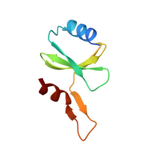
    DsbC and DsbG are periplasmic disulfide-bond isomerases, enzymes that facilitate the folding of secreted proteins with multiple disulfide bonds by catalyzing disulfide-bond rearrangement. Both enzymes also have in vitro chaperone activity. The crystal structures of these molecules are similar and both are V-shaped homodimeric modular structures. Each dimeric molecule contains two separate C-terminal thioredoxin-fold domains, joined by hinged helical "stalks" to a single N-terminal dimerization domain formed from the N-terminal 67 residues of each monomer. In this work, the crystal structures of the separate DsbC and DsbG dimerization domains have been determined at resolutions of 2.0 and 1.9 A, respectively. The two structures are both similar to the corresponding domains in the full-length molecules, showing that the dimerization domains fold independently of the catalytic portions of the full-length molecules. Localized structural differences between DsbC and DsbG were observed near the dimer interface and may be relevant to the different functions of the two enzymes.


  • Organizational Affiliation

    School of Biological Sciences, Auckland University, Auckland, New Zealand.


Macromolecules
Find similar proteins by:  (by identity cutoff)  |  3D Structure
Entity ID: 1
MoleculeChains Sequence LengthOrganismDetailsImage
THIOL DISULFIDE INTERCHANGE PROTEIN DSBC
A, B
75Escherichia coliMutation(s): 0 
UniProt
Find proteins for Q14F36 (Escherichia coli)
Explore Q14F36 
Go to UniProtKB:  Q14F36
Entity Groups  
Sequence Clusters30% Identity50% Identity70% Identity90% Identity95% Identity100% Identity
UniProt GroupQ14F36
Sequence Annotations
Expand
  • Reference Sequence
Small Molecules
Ligands 1 Unique
IDChains Name / Formula / InChI Key2D Diagram3D Interactions
SO4
Query on SO4

Download Ideal Coordinates CCD File 
C [auth B]SULFATE ION
O4 S
QAOWNCQODCNURD-UHFFFAOYSA-L
Experimental Data & Validation

Experimental Data

  • Method: X-RAY DIFFRACTION
  • Resolution: 2.00 Å
  • R-Value Free: 0.251 
  • R-Value Work: 0.226 
  • R-Value Observed: 0.228 
  • Space Group: P 65 2 2
Unit Cell:
Length ( Å )Angle ( ˚ )
a = 42.138α = 90
b = 42.138β = 90
c = 268.677γ = 120
Software Package:
Software NamePurpose
REFMACrefinement
DENZOdata reduction
SCALEPACKdata scaling
CCP4phasing

Structure Validation

View Full Validation Report


Entry History 

Deposition Data

Revision History  (Full details and data files)

  • Version 1.0: 2007-07-24
    Type: Initial release
  • Version 1.1: 2011-07-13
    Changes: Advisory, Refinement description, Version format compliance
  • Version 1.2: 2023-12-13
    Changes: Data collection, Database references, Other, Refinement description