


The crystal structure of SpoVG from Staphylococcus epidermidis ATCC 12228
Tan, K., Maltseva, N., Bargassa, M., Joachimiak, A.To be published.
Experimental Data Snapshot
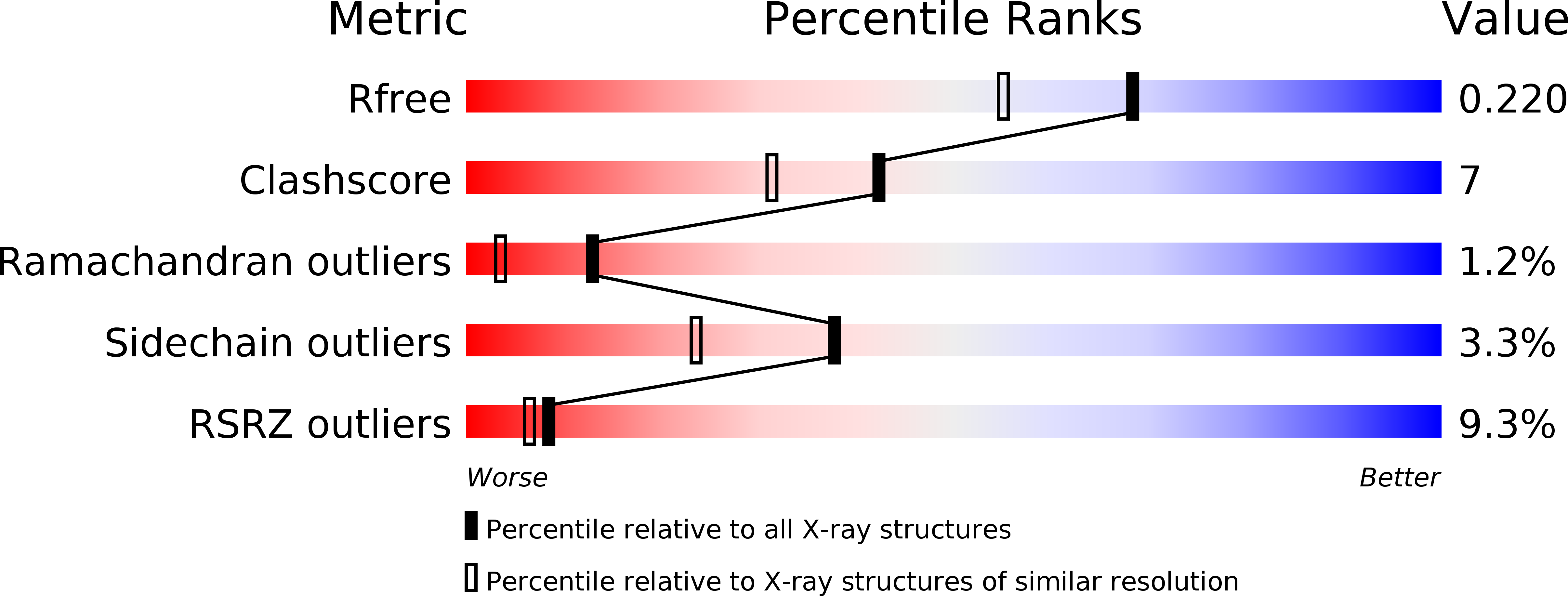
wwPDB Validation 3D Report Full Report
Entity ID: 1 | |||||
|---|---|---|---|---|---|
| Molecule | Chains | Sequence Length | Organism | Details | Image |
| Putative septation protein spoVG | 87 | Staphylococcus epidermidis ATCC 12228 | Mutation(s): 5 Gene Names: spoVG, SE_2285 |  | |
UniProt | |||||
Find proteins for Q8CML1 (Staphylococcus epidermidis (strain ATCC 12228 / FDA PCI 1200)) Explore Q8CML1 Go to UniProtKB: Q8CML1 | |||||
Entity Groups | |||||
| Sequence Clusters | 30% Identity50% Identity70% Identity90% Identity95% Identity100% Identity | ||||
| UniProt Group | Q8CML1 | ||||
Sequence AnnotationsExpand | |||||
| |||||
| Ligands 1 Unique | |||||
|---|---|---|---|---|---|
| ID | Chains | Name / Formula / InChI Key | 2D Diagram | 3D Interactions | |
| EDO Query on EDO | C [auth A], D [auth A], E [auth B], F [auth B], G [auth B] | 1,2-ETHANEDIOL C2 H6 O2 LYCAIKOWRPUZTN-UHFFFAOYSA-N |  | ||
| Modified Residues 1 Unique | |||||
|---|---|---|---|---|---|
| ID | Chains | Type | Formula | 2D Diagram | Parent |
| MSE Query on MSE | A, B | L-PEPTIDE LINKING | C5 H11 N O2 Se |  | MET |
| Length ( Å ) | Angle ( ˚ ) |
|---|---|
| a = 64.55 | α = 90 |
| b = 64.55 | β = 90 |
| c = 97.711 | γ = 90 |
| Software Name | Purpose |
|---|---|
| REFMAC | refinement |
| SBC-Collect | data collection |
| HKL-2000 | data reduction |
| HKL-2000 | data scaling |
| HKL-3000 | phasing |
| SHELXE | model building |
| SOLVE | phasing |
| RESOLVE | phasing |
| ARP/wARP | model building |
RCSB PDB (citation) is hosted by
RCSB PDB is a member of the

