2HUM

Crystal structure of T4 Lysozyme D72C synthetic dimer


Experimental Data Snapshot

  • Method: X-RAY DIFFRACTION
  • Resolution: 2.35 Å
  • R-Value Free: 0.304 
  • R-Value Work: 0.239 
  • R-Value Observed: 0.243 

wwPDB Validation   3D Report Full Report


This is version 1.4 of the entry. See complete history


Literature

An approach to crystallizing proteins by synthetic symmetrization.

Banatao, D.R.Cascio, D.Crowley, C.S.Fleissner, M.R.Tienson, H.L.Yeates, T.O.

(2006) Proc Natl Acad Sci U S A 103: 16230-16235

  • DOI: https://doi.org/10.1073/pnas.0607674103
  • Primary Citation of Related Structures:  
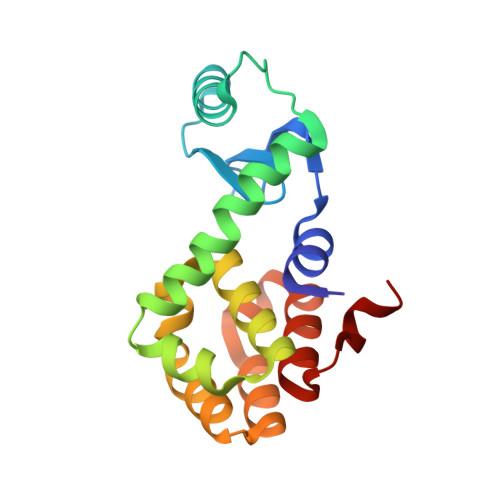
    2HUK, 2HUL, 2HUM

  • PubMed Abstract: 

    Previous studies of symmetry preferences in protein crystals suggest that symmetric proteins, such as homodimers, might crystallize more readily on average than asymmetric, monomeric proteins. Proteins that are naturally monomeric can be made homodimeric artificially by forming disulfide bonds between individual cysteine residues introduced by mutagenesis. Furthermore, by creating a variety of single-cysteine mutants, a series of distinct synthetic dimers can be generated for a given protein of interest, with each expected to gain advantage from its added symmetry and to exhibit a crystallization behavior distinct from the other constructs. This strategy was tested on phage T4 lysozyme, a protein whose crystallization as a monomer has been studied exhaustively. Experiments on three single-cysteine mutants, each prepared in dimeric form, yielded numerous novel crystal forms that cannot be realized by monomeric lysozyme. Six new crystal forms have been characterized. The results suggest that synthetic symmetrization may be a useful approach for enlarging the search space for crystallizing proteins.


  • Organizational Affiliation

    California Nanosystems Institute, University of California, Boyer Hall 259, P.O. Box 951570, Los Angeles, CA 90095-1570, USA.


Macromolecules
Find similar proteins by:  (by identity cutoff)  |  3D Structure
Entity ID: 1
MoleculeChains Sequence LengthOrganismDetailsImage
Lysozyme
A, B
164Tequatrovirus T4Mutation(s): 3 
Gene Names: E
EC: 3.2.1.17
UniProt
Find proteins for P00720 (Enterobacteria phage T4)
Explore P00720 
Go to UniProtKB:  P00720
Entity Groups  
Sequence Clusters30% Identity50% Identity70% Identity90% Identity95% Identity100% Identity
UniProt GroupP00720
Sequence Annotations
Expand
  • Reference Sequence
Small Molecules
Ligands 1 Unique
IDChains Name / Formula / InChI Key2D Diagram3D Interactions
TRS
Query on TRS

Download Ideal Coordinates CCD File 
C [auth A]2-AMINO-2-HYDROXYMETHYL-PROPANE-1,3-DIOL
C4 H12 N O3
LENZDBCJOHFCAS-UHFFFAOYSA-O
Experimental Data & Validation

Experimental Data

  • Method: X-RAY DIFFRACTION
  • Resolution: 2.35 Å
  • R-Value Free: 0.304 
  • R-Value Work: 0.239 
  • R-Value Observed: 0.243 
  • Space Group: P 1 21 1
Unit Cell:
Length ( Å )Angle ( ˚ )
a = 55.306α = 90
b = 55.907β = 108.13
c = 80.292γ = 90
Software Package:
Software NamePurpose
REFMACrefinement
DENZOdata reduction
SCALEPACKdata scaling
PHASERphasing

Structure Validation

View Full Validation Report


Entry History 

Deposition Data

Revision History  (Full details and data files)

  • Version 1.0: 2006-10-17
    Type: Initial release
  • Version 1.1: 2008-05-01
    Changes: Version format compliance
  • Version 1.2: 2011-07-13
    Changes: Version format compliance
  • Version 1.3: 2021-10-20
    Changes: Database references, Derived calculations
  • Version 1.4: 2023-08-30
    Changes: Data collection, Refinement description