Structural and Biophysical Characterization of the EphB4-EphrinB2 Protein-Protein Interaction and Receptor Specificity.
Chrencik, J.E., Brooun, A., Kraus, M.L., Recht, M.I., Kolatkar, A.R., Han, G.W., Seifert, J.M., Widmer, H., Auer, M., Kuhn, P.(2006) J Biol Chem 281: 28185-28192
- PubMed: 16867992
- DOI: https://doi.org/10.1074/jbc.M605766200
- Primary Citation of Related Structures:
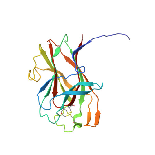
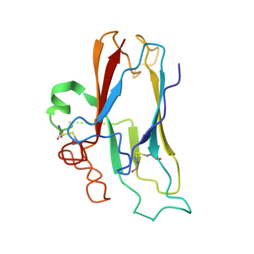
2HLE - PubMed Abstract:
Increasing evidence implicates the interaction of the EphB4 receptor with its preferred ligand, ephrinB2, in pathological forms of angiogenesis and in tumorigenesis. To identify the molecular determinants of the unique specificity of EphB4 for ephrinB2, we determined the crystal structure of the ligand binding domain of EphB4 in complex with the extracellular domain of ephrinB2. This structural analysis suggested that one amino acid, Leu-95, plays a particularly important role in defining the structural features that confer the ligand selectivity of EphB4. Indeed, all other Eph receptors, which promiscuously bind many ephrins, have a conserved arginine at the position corresponding to Leu-95 of EphB4. We have also found that amino acid changes in the EphB4 ligand binding cavity, designed based on comparison with the crystal structure of the more promiscuous EphB2 receptor, yield EphB4 variants with altered binding affinity for ephrinB2 and an antagonistic peptide. Isothermal titration calorimetry experiments with an EphB4 Leu-95 to arginine mutant confirmed the importance of this amino acid in conferring high affinity binding to both ephrinB2 and the antagonistic peptide ligand. Isothermal titration calorimetry measurements also revealed an interesting thermodynamic discrepancy between ephrinB2 binding, which is an entropically driven process, and peptide binding, which is an enthalpically driven process. These results provide critical information on the EphB4*ephrinB2 protein interfaces and their mode of interaction, which will facilitate development of small molecule compounds inhibiting the EphB4*ephrinB2 interaction as novel cancer therapeutics.
Organizational Affiliation:
Department of Cellular Biology, The Scripps Research Institute, La Jolla, California 92037, USA.