2HA0

Crystal structure of mouse acetylcholinesterase complexed with 4-ketoamyltrimethylammonium


Experimental Data Snapshot

  • Method: X-RAY DIFFRACTION
  • Resolution: 2.20 Å
  • R-Value Free: 0.219 
  • R-Value Work: 0.182 
  • R-Value Observed: 0.183 

wwPDB Validation   3D Report Full Report


Ligand Structure Quality Assessment 


This is version 3.1 of the entry. See complete history


Literature

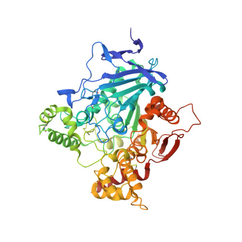
Substrate and product trafficking through the active center gorge of acetylcholinesterase analyzed by crystallography and equilibrium binding

Bourne, Y.Radic, Z.Sulzenbacher, G.Kim, E.Taylor, P.Marchot, P.

(2006) J Biol Chem 281: 29256-29267

  • DOI: https://doi.org/10.1074/jbc.M603018200
  • Primary Citation of Related Structures:  
    2H9Y, 2HA0, 2HA2, 2HA3, 2HA4, 2HA5, 2HA6, 2HA7

  • PubMed Abstract: 

    Hydrolysis of acetylcholine catalyzed by acetylcholinesterase (AChE), one of the most efficient enzymes in nature, occurs at the base of a deep and narrow active center gorge. At the entrance of the gorge, the peripheral anionic site provides a binding locus for allosteric ligands, including substrates. To date, no structural information on substrate entry to the active center from the peripheral site of AChE or its subsequent egress has been reported. Complementary crystal structures of mouse AChE and an inactive mouse AChE mutant with a substituted catalytic serine (S203A), in various complexes with four substrates (acetylcholine, acetylthiocholine, succinyldicholine, and butyrylthiocholine), two non-hydrolyzable substrate analogues (m-(N,N,N-trimethylammonio)-trifluoroacetophenone and 4-ketoamyltrimethylammonium), and one reaction product (choline) were solved in the 2.05-2.65-A resolution range. These structures, supported by binding and inhibition data obtained on the same complexes, reveal the successive positions and orientations of the substrates bound to the peripheral site and proceeding within the gorge toward the active site, the conformations of the presumed transition state for acylation and the acyl-enzyme intermediate, and the positions and orientations of the dissociating and egressing products. Moreover, the structures of the AChE mutant in complexes with acetylthiocholine and succinyldicholine reveal additional substrate binding sites on the enzyme surface, distal to the gorge entry. Hence, we provide a comprehensive set of structural snapshots of the steps leading to the intermediates of catalysis and the potential regulation by substrate binding to various allosteric sites at the enzyme surface.


  • Organizational Affiliation

    Ingénierie des Protéines, CNRS FRE-2738, Institut Fédératif de Recherche Jean Roche, Université dela Méditerranée, Faculté d eMédecine Secteur Nord, F-13916 Marseille Cedex 20, France.


Macromolecules
Find similar proteins by:  (by identity cutoff)  |  3D Structure
Entity ID: 1
MoleculeChains Sequence LengthOrganismDetailsImage
Acetylcholinesterase
A, B
543Mus musculusMutation(s): 0 
EC: 3.1.1.7
UniProt & NIH Common Fund Data Resources
Find proteins for P21836 (Mus musculus)
Explore P21836 
Go to UniProtKB:  P21836
IMPC:  MGI:87876
Entity Groups  
Sequence Clusters30% Identity50% Identity70% Identity90% Identity95% Identity100% Identity
UniProt GroupP21836
Sequence Annotations
Expand
  • Reference Sequence
Oligosaccharides

Help

Entity ID: 2
MoleculeChains Length2D Diagram Glycosylation3D Interactions
alpha-L-fucopyranose-(1-6)-2-acetamido-2-deoxy-beta-D-glucopyranose
C
2N-Glycosylation
Glycosylation Resources
GlyTouCan:  G86851RC
GlyCosmos:  G86851RC
GlyGen:  G86851RC
Small Molecules
Ligands 5 Unique
IDChains Name / Formula / InChI Key2D Diagram3D Interactions
P6G
Query on P6G

Download Ideal Coordinates CCD File 
N [auth A]HEXAETHYLENE GLYCOL
C12 H26 O7
IIRDTKBZINWQAW-UHFFFAOYSA-N
NAG
Query on NAG

Download Ideal Coordinates CCD File 
D [auth A],
O [auth B]
2-acetamido-2-deoxy-beta-D-glucopyranose
C8 H15 N O6
OVRNDRQMDRJTHS-FMDGEEDCSA-N
NWA
Query on NWA

Download Ideal Coordinates CCD File 
L [auth A],
W [auth B]
4,4-DIHYDROXY-N,N,N-TRIMETHYLPENTAN-1-AMINIUM
C8 H20 N O2
WEYSSHXEYINXQE-UHFFFAOYSA-N
CHH
Query on CHH

Download Ideal Coordinates CCD File 
M [auth A],
X [auth B]
N,N,N-TRIMETHYL-4-OXOPENTAN-1-AMINIUM
C8 H18 N O
UKCYTFTWLWVZSO-UHFFFAOYSA-N
IOD
Query on IOD

Download Ideal Coordinates CCD File 
E [auth A]
F [auth A]
G [auth A]
H [auth A]
I [auth A]
E [auth A],
F [auth A],
G [auth A],
H [auth A],
I [auth A],
J [auth A],
K [auth A],
P [auth B],
Q [auth B],
R [auth B],
S [auth B],
T [auth B],
U [auth B],
V [auth B]
IODIDE ION
I
XMBWDFGMSWQBCA-UHFFFAOYSA-M
Binding Affinity Annotations 
IDSourceBinding Affinity
CHH BindingDB:  2HA0 Ki: 2.30e+4 (nM) from 1 assay(s)
IC50: 8.47e+4 (nM) from 1 assay(s)
Binding MOAD:  2HA0 Kd: 3.70e+4 (nM) from 1 assay(s)
NWA PDBBind:  2HA0 Kd: 3.70e+4 (nM) from 1 assay(s)
Experimental Data & Validation

Experimental Data

  • Method: X-RAY DIFFRACTION
  • Resolution: 2.20 Å
  • R-Value Free: 0.219 
  • R-Value Work: 0.182 
  • R-Value Observed: 0.183 
  • Space Group: P 21 21 21
Unit Cell:
Length ( Å )Angle ( ˚ )
a = 79.228α = 90
b = 111.795β = 90
c = 226.94γ = 90
Software Package:
Software NamePurpose
REFMACrefinement
ADSCdata collection
DENZOdata reduction
SCALEPACKdata scaling

Structure Validation

View Full Validation Report


Ligand Structure Quality Assessment 


Entry History 

Deposition Data

Revision History  (Full details and data files)

  • Version 1.0: 2006-07-18
    Type: Initial release
  • Version 1.1: 2008-05-01
    Changes: Version format compliance
  • Version 1.2: 2011-07-13
    Changes: Advisory, Version format compliance
  • Version 1.3: 2011-10-05
    Changes: Derived calculations
  • Version 2.0: 2020-07-29
    Type: Remediation
    Reason: Carbohydrate remediation
    Changes: Advisory, Atomic model, Data collection, Derived calculations, Structure summary
  • Version 3.0: 2021-03-10
    Changes: Advisory, Atomic model, Data collection, Derived calculations, Structure summary
  • Version 3.1: 2023-10-25
    Changes: Data collection, Database references, Refinement description