The Crystal Structure of GCAP3 Suggests Molecular Mechanism of GCAP-linked Cone Dystrophies.
Stephen, R., Palczewski, K., Sousa, M.C.(2006) J Mol Biol 359: 266-275
- PubMed: 16626734
- DOI: https://doi.org/10.1016/j.jmb.2006.03.042
- Primary Citation of Related Structures:
2GGZ - PubMed Abstract:
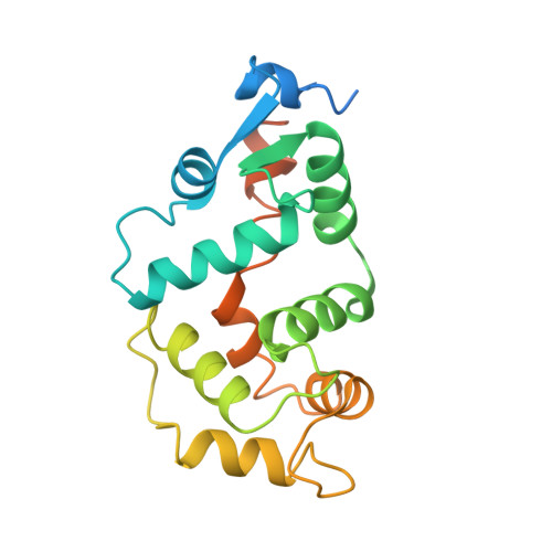
Absorption of light by visual pigments initiates the phototransduction pathway that results in degradation of the intracellular pool of cyclic-GMP (cGMP). This hydrolysis promotes the closing of cGMP-gated cation channels and consequent hyperpolarization of rod and cone photoreceptor cell membranes. Guanylate cyclase-activating proteins (GCAPs) are a family of proteins that regulate retinal guanylate cyclase (GC) activity in a Ca2+-dependent manner. At high [Ca2+], typical of the dark-adapted state (approximately 500 nM), GCAPs inhibit retinal GCs. At the low [Ca2+] (approximately 50 nM) that occurs after the closing of cGMP-gated channels, GCAPs activate retinal GCs to replenish dark-state cGMP levels. Here, we report the crystal structure of unmyristoylated human GCAP3 with Ca2+ bound. GCAP3 is an EF-hand Ca2+-binding protein with Ca2+ bound to EF2, 3 and 4, while Ca2+ binding to EF-hand 1 is disabled. GCAP3 contains two domains with the EF-hand motifs arranged in a tandem array similar to GCAP2 and members of the recoverin subfamily of Ca2+-binding proteins. Residues not involved in Ca2+ binding, but conserved in all GCAPs, cluster around EF1 in the N-terminal domain and may represent the interface with GCs. Five point mutations in the closely related GCAP1 have been linked to the etiology of cone dystrophies. These residues are conserved in GCAP3 and the structure suggests important roles for these amino acids. We present a homology model of GCAP1 based on GCAP3 that offers insight into the molecular mechanism underlying the autosomal dominant cone dystrophies produced by GCAP1 mutations.
Organizational Affiliation:
Department of Chemistry and Biochemistry, University of Colorado at Boulder, Boulder, CO 80309, USA.