2FXU

X-ray Structure of Bistramide A- Actin Complex at 1.35 A resolution.


Experimental Data Snapshot

  • Method: X-RAY DIFFRACTION
  • Resolution: 1.35 Å
  • R-Value Free: 0.201 
  • R-Value Work: 0.174 
  • R-Value Observed: 0.176 

wwPDB Validation   3D Report Full Report


Ligand Structure Quality Assessment 


This is version 1.3 of the entry. See complete history


Literature

Structure of bistramide a-actin complex at a 1.35 A resolution

Rizvi, S.A.Tereshko, V.Kossiakoff, A.A.Kozmin, S.A.

(2006) J Am Chem Soc 128: 3882-3883

  • DOI: https://doi.org/10.1021/ja058319c
  • Primary Citation of Related Structures:  
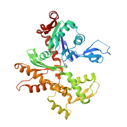
    2FXU

  • PubMed Abstract: 

    Bistramide A is a highly potent antiproliferative marine natural product from Lissoclinum bistratum. We have previously established actin as the primary cellular receptor of bistramide A. We report herein the X-ray structure of bistramide A bound to monomeric actin at a resolution of 1.35 A. The most notable aspect of the bistramide A-actin structure is an extensive hydrogen-bonding network established upon a deep penetration of the central segment of bistramide A into the actin-binding cleft between subdomains 1 and 3. The structure presents the first insight into the observed ability of bistramide A to modulate G-actin polymerization. The structural information combined with our ability to chemically modify the bistramide framework provides the basis for rational development of a series of new synthetic analogues as useful probes for studying actin cytoskeleton and as potential therapeutic leads.


  • Organizational Affiliation

    Department of Chemistry, University of Chicago, Chicago, Illinois 60637, USA.


Macromolecules
Find similar proteins by:  (by identity cutoff)  |  3D Structure
Entity ID: 1
MoleculeChains Sequence LengthOrganismDetailsImage
Actin, alpha skeletal muscle375Oryctolagus cuniculusMutation(s): 1 
UniProt
Find proteins for P68135 (Oryctolagus cuniculus)
Explore P68135 
Go to UniProtKB:  P68135
Entity Groups  
Sequence Clusters30% Identity50% Identity70% Identity90% Identity95% Identity100% Identity
UniProt GroupP68135
Sequence Annotations
Expand
  • Reference Sequence
Small Molecules
Modified Residues  1 Unique
IDChains TypeFormula2D DiagramParent
HIC
Query on HIC
A
L-PEPTIDE LINKINGC7 H11 N3 O2HIS
Binding Affinity Annotations 
IDSourceBinding Affinity
BID PDBBind:  2FXU Kd: 7 (nM) from 1 assay(s)
Binding MOAD:  2FXU Kd: 7 (nM) from 1 assay(s)
Experimental Data & Validation

Experimental Data

  • Method: X-RAY DIFFRACTION
  • Resolution: 1.35 Å
  • R-Value Free: 0.201 
  • R-Value Work: 0.174 
  • R-Value Observed: 0.176 
  • Space Group: C 1 2 1
Unit Cell:
Length ( Å )Angle ( ˚ )
a = 60.125α = 90
b = 56.513β = 94.56
c = 101.652γ = 90
Software Package:
Software NamePurpose
REFMACrefinement
HKL-2000data reduction
SCALEPACKdata scaling
MOLREPphasing

Structure Validation

View Full Validation Report


Ligand Structure Quality Assessment 


Entry History 

Deposition Data

Revision History  (Full details and data files)

  • Version 1.0: 2006-03-07
    Type: Initial release
  • Version 1.1: 2008-05-01
    Changes: Version format compliance
  • Version 1.2: 2011-07-13
    Changes: Advisory, Refinement description, Version format compliance
  • Version 1.3: 2023-08-30
    Changes: Data collection, Database references, Derived calculations, Refinement description, Structure summary