2F86

The Association Domain of C. elegans CaMKII


Experimental Data Snapshot

  • Method: X-RAY DIFFRACTION
  • Resolution: 2.64 Å
  • R-Value Free: 0.301 
  • R-Value Work: 0.248 
  • R-Value Observed: 0.248 

wwPDB Validation   3D Report Full Report


This is version 1.5 of the entry. See complete history


Literature

Oligomerization states of the association domain and the holoenyzme of Ca2+/CaM kinase II.

Rosenberg, O.S.Deindl, S.Comolli, L.R.Hoelz, A.Downing, K.H.Nairn, A.C.Kuriyan, J.

(2006) FEBS J 273: 682-694

  • DOI: https://doi.org/10.1111/j.1742-4658.2005.05088.x
  • Primary Citation of Related Structures:  
    2F86

  • PubMed Abstract: 

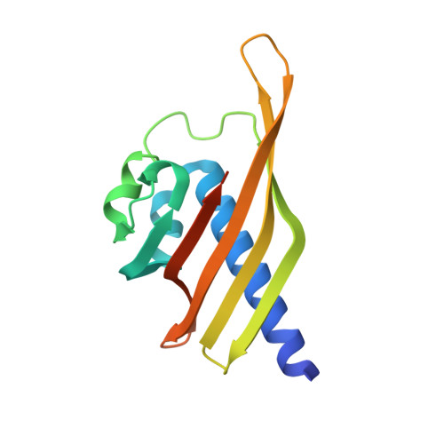
    Ca2+/calmodulin activated protein kinase II (CaMKII) is an oligomeric protein kinase with a unique holoenyzme architecture. The subunits of CaMKII are bound together into the holoenzyme by the association domain, a C-terminal region of approximately 140 residues in the CaMKII polypeptide. Single particle analyses of electron micrographs have suggested previously that the holoenyzme forms a dodecamer that contains two stacked 6-fold symmetric rings. In contrast, a recent crystal structure of the isolated association domain of mouse CaMKIIalpha has revealed a tetradecameric assembly with two stacked 7-fold symmetric rings. In this study, we have determined the crystal structure of the Caenorhabditis elegans CaMKII association domain and it too forms a tetradecamer. We also show by electron microscopy that in its fully assembled form the CaMKII holoenzyme is a dodecamer but without the kinase domains, either from expression of the isolated association domain in bacteria or following their removal by proteolysis, the association domains form a tetradecamer. We speculate that the holoenzyme is held in its 6-fold symmetric state by the interactions of the N-terminal approximately 1-335 residues and that the removal of this region allows the association domain to convert into a more stable 7-fold symmetric form.


  • Organizational Affiliation

    Department of Molecular and Cell Biology, University of California, Berkeley, CA 94720-3202, USA.


Macromolecules
Find similar proteins by:  (by identity cutoff)  |  3D Structure
Entity ID: 1
MoleculeChains Sequence LengthOrganismDetailsImage
Hypothetical protein K11E8.1d143Caenorhabditis elegansMutation(s): 0 
Gene Names: unc-43K11E8.1
EC: 2.7.11.17
UniProt
Find proteins for O62305 (Caenorhabditis elegans)
Explore O62305 
Go to UniProtKB:  O62305
Entity Groups  
Sequence Clusters30% Identity50% Identity70% Identity90% Identity95% Identity100% Identity
UniProt GroupO62305
Sequence Annotations
Expand
  • Reference Sequence
Experimental Data & Validation

Experimental Data

  • Method: X-RAY DIFFRACTION
  • Resolution: 2.64 Å
  • R-Value Free: 0.301 
  • R-Value Work: 0.248 
  • R-Value Observed: 0.248 
  • Space Group: C 2 2 21
Unit Cell:
Length ( Å )Angle ( ˚ )
a = 70.87α = 90
b = 186.934β = 90
c = 182.934γ = 90
Software Package:
Software NamePurpose
CNSrefinement
MADNESSdata reduction
SCALEPACKdata scaling
PHASERphasing

Structure Validation

View Full Validation Report


Entry History 

Deposition Data

Revision History  (Full details and data files)

  • Version 1.0: 2006-02-07
    Type: Initial release
  • Version 1.1: 2008-05-01
    Changes: Version format compliance
  • Version 1.2: 2011-07-13
    Changes: Derived calculations, Version format compliance
  • Version 1.3: 2017-06-28
    Changes: Database references, Source and taxonomy, Structure summary
  • Version 1.4: 2017-10-18
    Changes: Refinement description
  • Version 1.5: 2023-08-30
    Changes: Data collection, Database references, Refinement description