2F42

dimerization and U-box domains of Zebrafish C-terminal of HSP70 interacting protein


Experimental Data Snapshot

  • Method: X-RAY DIFFRACTION
  • Resolution: 2.50 Å
  • R-Value Free: 0.285 
  • R-Value Work: 0.266 
  • R-Value Observed: 0.266 

wwPDB Validation   3D Report Full Report


This is version 1.3 of the entry. See complete history


Literature

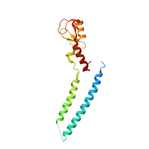
Structure and Interactions of the Helical and U-Box Domains of CHIP, the C Terminus of HSP70 Interacting Protein.

Xu, Z.Devlin, K.I.Ford, M.G.Nix, J.C.Qin, J.Misra, S.

(2006) Biochemistry 45: 4749-4759

  • DOI: https://doi.org/10.1021/bi0601508
  • Primary Citation of Related Structures:  
    2F42

  • PubMed Abstract: 

    The heat-shock proteins Hsp70 and Hsp90 play a crucial role in regulating protein quality control both by refolding and by preventing the aggregation of misfolded proteins. It has recently been shown that Hsp70 and Hsp90 act not only in protein refolding but also cooperate with the C terminus of Hsp70 interacting protein (CHIP), a multidomain ubiquitin ligase, to mediate the degradation of unfolded proteins. We present the crystal structure of the helical linker domain and U-box domain of zebrafish CHIP (DrCHIP-HU). The structure of DrCHIP-HU shows a symmetric homodimer. The conformation of the helical linker domains and the relative positions of the helical and U-box domains differ substantially in DrCHIP-HU from those in a recently published structure of an asymmetric dimer of mammalian (mouse) CHIP. We used an in vitro ubiquitination assay to identify residues, located on two long loops and a central alpha helix of the CHIP U-box domain, that are important for interacting with the ubiquitin-conjugating enzyme UbcH5b. In addition, we used NMR spectroscopy to define a complementary interaction surface located on the N-terminal alpha helix and the L4 and L7 loops of UbcH5b. Our results provide insights into conformational variability in the domain arrangement of CHIP and into U-box-mediated recruitment of UbcH5b for the ubiquitination of Hsp70 and Hsp90 substrates.


  • Organizational Affiliation

    Department of Molecular Cardiology, The Cleveland Clinic Foundation, Cleveland, Ohio 44195, USA.


Macromolecules
Find similar proteins by:  (by identity cutoff)  |  3D Structure
Entity ID: 1
MoleculeChains Sequence LengthOrganismDetailsImage
STIP1 homology and U-box containing protein 1179Danio rerioMutation(s): 0 
Gene Names: stub1 (amino acids 112-284)
UniProt
Find proteins for F6NGW2 (Danio rerio)
Explore F6NGW2 
Go to UniProtKB:  F6NGW2
Entity Groups  
Sequence Clusters30% Identity50% Identity70% Identity90% Identity95% Identity100% Identity
UniProt GroupF6NGW2
Sequence Annotations
Expand
  • Reference Sequence
Small Molecules
Ligands 1 Unique
IDChains Name / Formula / InChI Key2D Diagram3D Interactions
CL
Query on CL

Download Ideal Coordinates CCD File 
B [auth A]CHLORIDE ION
Cl
VEXZGXHMUGYJMC-UHFFFAOYSA-M
Experimental Data & Validation

Experimental Data

  • Method: X-RAY DIFFRACTION
  • Resolution: 2.50 Å
  • R-Value Free: 0.285 
  • R-Value Work: 0.266 
  • R-Value Observed: 0.266 
  • Space Group: P 65 2 2
Unit Cell:
Length ( Å )Angle ( ˚ )
a = 100.499α = 90
b = 100.499β = 90
c = 74.311γ = 120
Software Package:
Software NamePurpose
d*TREKdata scaling
SOLVEphasing
RESOLVEphasing
CNSrefinement
PDB_EXTRACTdata extraction
d*TREKdata reduction

Structure Validation

View Full Validation Report


Entry History 

Deposition Data

Revision History  (Full details and data files)

  • Version 1.0: 2006-05-02
    Type: Initial release
  • Version 1.1: 2008-05-01
    Changes: Version format compliance
  • Version 1.2: 2011-07-13
    Changes: Derived calculations, Version format compliance
  • Version 1.3: 2024-02-14
    Changes: Data collection, Database references, Derived calculations