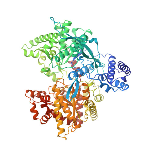
The crystal structure of the Escherichia coli maltodextrin phosphorylase-acarbose complex.
O'Reilly, M., Watson, K.A., Johnson, L.N.(1999) Biochemistry 38: 5337-5345
- PubMed: 10220320
- DOI: https://doi.org/10.1021/bi9828573
- Primary Citation of Related Structures:
2ECP - PubMed Abstract:
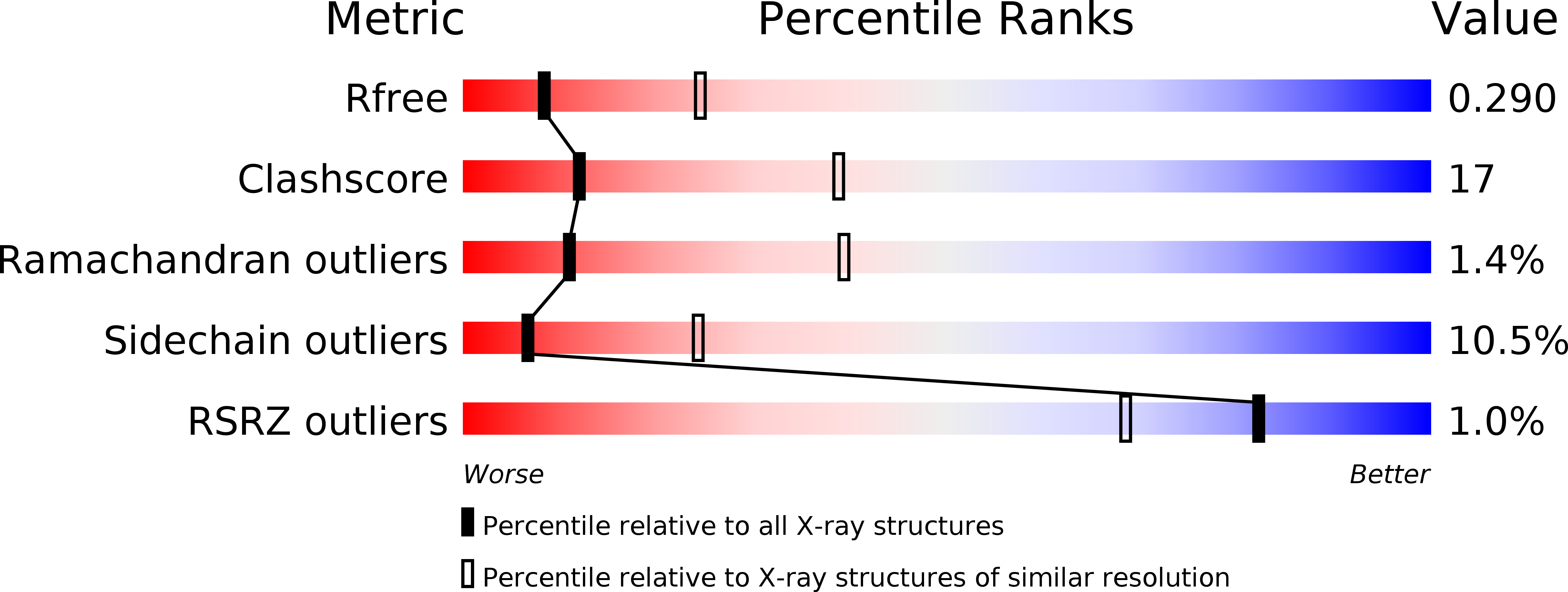
Acarbose is a naturally occurring pseudo-tetrasaccharide. It has been used in conjunction with other drugs in the treatment of diabetes where it acts as an inhibitor of intestinal glucosidases. To probe the interactions of acarbose with other carbohydrate recognition enzymes, the crystal structure of E. coli maltodextrin phosphorylase (MalP) complexed with acarbose has been determined at 2.95 A resolution and refined to crystallographic R-values of R (Rfree) = 0.241 (0.293), respectively. Acarbose adopts a conformation that is close to its major minimum free energy conformation in the MalP-acarbose structure. The acarviosine moiety of acarbose occupies sub-sites +1 and +2 and the disaccharide sub-sites +3 and +4. (The site of phosphorolysis is between sub-sites -1 and +1.) This is the first identification of sub-sites +3 and +4 of MalP. Interactions of the glucosyl residues in sub-sites +2 and +4 are dominated by carbohydrate stacking interactions with tyrosine residues. These tyrosines (Tyr280 and Tyr613, respectively, in the rabbit muscle phosphorylase numbering scheme) are conserved in all species of phosphorylase. A glycerol molecule from the cryoprotectant occupies sub-site -1. The identification of four oligosaccharide sub-sites, that extend from the interior of the phosphorylase close to the catalytic site to the exterior surface of MalP, provides a structural rationalization of the substrate selectivity of MalP for a pentasaccharide substrate. Crystallographic binding studies of acarbose with amylases, glucoamylases, and glycosyltranferases and NMR studies of acarbose in solution have shown that acarbose can adopt two different conformations. This flexibility allows acarbose to target a number of different enzymes. The two alternative conformations of acarbose when bound to different carbohydrate enzymes are discussed.
Organizational Affiliation:
Laboratory of Molecular Biophysics, Oxford Centre for Molecular Sciences, Department of Biochemistry, University of Oxford, U.K.