2E0I

Crystal structure of archaeal photolyase from Sulfolobus tokodaii with two FAD molecules: Implication of a novel light-harvesting cofactor


Experimental Data Snapshot

  • Method: X-RAY DIFFRACTION
  • Resolution: 2.80 Å
  • R-Value Free: 0.247 
  • R-Value Work: 0.223 

Starting Model: experimental
View more details

wwPDB Validation   3D Report Full Report


This is version 1.3 of the entry. See complete history


Literature

Crystal Structure of Archaeal Photolyase from Sulfolobus tokodaii with Two FAD Molecules: Implication of a Novel Light-harvesting Cofactor

Fujihashi, M.Numoto, N.Kobayashi, Y.Mizushima, A.Tsujimura, M.Nakamura, A.Kawarabayasi, Y.Miki, K.

(2007) J Mol Biol 365: 903-910

  • DOI: https://doi.org/10.1016/j.jmb.2006.10.012
  • Primary Citation of Related Structures:  
    2E0I

  • PubMed Abstract: 

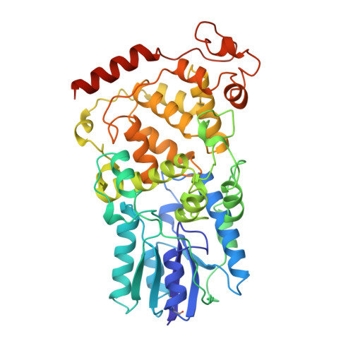
    UV exposure of DNA molecules induces serious DNA lesions. The cyclobutane pyrimidine dimer (CPD) photolyase repairs CPD-type - lesions by using the energy of visible light. Two chromophores for different roles have been found in this enzyme family; one catalyzes the CPD repair reaction and the other works as an antenna pigment that harvests photon energy. The catalytic cofactor of all known photolyases is FAD, whereas several light-harvesting cofactors are found. Currently, 5,10-methenyltetrahydrofolate (MTHF), 8-hydroxy-5-deaza-riboflavin (8-HDF) and FMN are the known light-harvesting cofactors, and some photolyases lack the chromophore. Three crystal structures of photolyases from Escherichia coli (Ec-photolyase), Anacystis nidulans (An-photolyase), and Thermus thermophilus (Tt-photolyase) have been determined; however, no archaeal photolyase structure is available. A similarity search of archaeal genomic data indicated the presence of a homologous gene, ST0889, on Sulfolobus tokodaii strain7. An enzymatic assay reveals that ST0889 encodes photolyase from S. tokodaii (St-photolyase). We have determined the crystal structure of the St-photolyase protein to confirm its structural features and to investigate the mechanism of the archaeal DNA repair system with light energy. The crystal structure of the St-photolyase is superimposed very well on the three known photolyases including the catalytic cofactor FAD. Surprisingly, another FAD molecule is found at the position of the light-harvesting cofactor. This second FAD molecule is well accommodated in the crystal structure, suggesting that FAD works as a novel light-harvesting cofactor of photolyase. In addition, two of the four CPD recognition residues in the crystal structure of An-photolyase are not found in St-photolyase, which might utilize a different mechanism to recognize the CPD from that of An-photolyase.


  • Organizational Affiliation

    Department of Chemistry, Graduate School of Science, Kyoto University, Sakyo-ku, Kyoto 606-8502, Japan.


Macromolecules
Find similar proteins by:  (by identity cutoff)  |  3D Structure
Entity ID: 1
MoleculeChains Sequence LengthOrganismDetailsImage
432aa long hypothetical deoxyribodipyrimidine photolyase
A, B, C, D
440Sulfurisphaera tokodaii str. 7Mutation(s): 0 
Gene Names: ST0889
EC: 4.1.99.3
UniProt
Find proteins for F9VNB1 (Sulfurisphaera tokodaii (strain DSM 16993 / JCM 10545 / NBRC 100140 / 7))
Explore F9VNB1 
Go to UniProtKB:  F9VNB1
Entity Groups  
Sequence Clusters30% Identity50% Identity70% Identity90% Identity95% Identity100% Identity
UniProt GroupF9VNB1
Sequence Annotations
Expand
  • Reference Sequence
Small Molecules
Ligands 2 Unique
IDChains Name / Formula / InChI Key2D Diagram3D Interactions
FAD
Query on FAD

Download Ideal Coordinates CCD File 
E [auth A]
F [auth A]
I [auth B]
J [auth B]
L [auth C]
E [auth A],
F [auth A],
I [auth B],
J [auth B],
L [auth C],
M [auth C],
P [auth D],
Q [auth D]
FLAVIN-ADENINE DINUCLEOTIDE
C27 H33 N9 O15 P2
VWWQXMAJTJZDQX-UYBVJOGSSA-N
MPD
Query on MPD

Download Ideal Coordinates CCD File 
G [auth A]
H [auth A]
K [auth B]
N [auth C]
O [auth C]
G [auth A],
H [auth A],
K [auth B],
N [auth C],
O [auth C],
R [auth D]
(4S)-2-METHYL-2,4-PENTANEDIOL
C6 H14 O2
SVTBMSDMJJWYQN-YFKPBYRVSA-N
Experimental Data & Validation

Experimental Data

  • Method: X-RAY DIFFRACTION
  • Resolution: 2.80 Å
  • R-Value Free: 0.247 
  • R-Value Work: 0.223 
  • Space Group: P 1 21 1
Unit Cell:
Length ( Å )Angle ( ˚ )
a = 79.875α = 90
b = 114.715β = 91.49
c = 167.684γ = 90
Software Package:
Software NamePurpose
CNSrefinement
HKL-2000data collection
HKL-2000data reduction
SCALEPACKdata scaling
MOLREPphasing

Structure Validation

View Full Validation Report


Entry History 

Deposition Data

Revision History  (Full details and data files)

  • Version 1.0: 2006-11-28
    Type: Initial release
  • Version 1.1: 2008-04-30
    Changes: Version format compliance
  • Version 1.2: 2011-07-13
    Changes: Source and taxonomy, Version format compliance
  • Version 1.3: 2023-10-25
    Changes: Data collection, Database references, Derived calculations, Refinement description