2DZ7

DNA Octaplex Formation with an I-Motif of A-Quartets: The Revised Crystal Structure of d(GCGAAAGC)


Experimental Data Snapshot

  • Method: X-RAY DIFFRACTION
  • Resolution: 1.60 Å
  • R-Value Free: 0.211 
  • R-Value Work: 0.190 
  • R-Value Observed: 0.190 

wwPDB Validation   3D Report Full Report


This is version 1.2 of the entry. See complete history


Literature

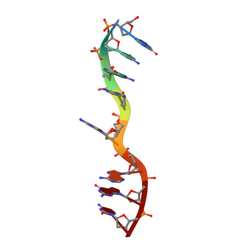
DNA octaplex formation with an I-motif of water-mediated A-quartets: reinterpretation of the crystal structure of d(GCGAAAGC)

Sato, Y.Mitomi, K.Sunami, T.Kondo, J.Takenaka, A.

(2006) J Biochem 140: 759-762

  • DOI: https://doi.org/10.1093/jb/mvj213
  • Primary Citation of Related Structures:  
    2DZ7

  • PubMed Abstract: 

    The crystal structure of the tetragonal form of d(gcGAAAgc) has been revised and reasonably refined including the disordered residues. The two DNA strands form a base-intercalated duplex, and the four duplexes are assembled according to the crystallographic 222 symmetry to form an octaplex. In the central region, the eight strands are associated by I-motif of double A-quartets. Furthermore, eight hydrated-magnesium cations link the four duplexes to support the octaplex formation. Based on these structural features, a proposal that folding of d(GAAA)n, found in the non-coding region of genomes, into an octaplex can induce slippage during replication to facilitate length polymorphism is presented.


  • Organizational Affiliation

    Graduate School of Bioscience and Biotechnology, Tokyo Institute of Technology, 4259 Nagatsuda, Midori-ku, Yokohama 226-850.


Macromolecules

Find similar nucleic acids by:  Sequence   |   3D Structure  

Entity ID: 1
MoleculeChains LengthOrganismImage
DNA (5'-D(*DGP*DCP*DGP*DAP*DAP*DAP*DGP*DC)-3')8N/A
Sequence Annotations
Expand
  • Reference Sequence
Small Molecules
Ligands 1 Unique
IDChains Name / Formula / InChI Key2D Diagram3D Interactions
MG
Query on MG

Download Ideal Coordinates CCD File 
B [auth A],
C [auth A]
MAGNESIUM ION
Mg
JLVVSXFLKOJNIY-UHFFFAOYSA-N
Experimental Data & Validation

Experimental Data

  • Method: X-RAY DIFFRACTION
  • Resolution: 1.60 Å
  • R-Value Free: 0.211 
  • R-Value Work: 0.190 
  • R-Value Observed: 0.190 
  • Space Group: I 4 2 2
Unit Cell:
Length ( Å )Angle ( ˚ )
a = 36.909α = 90
b = 36.909β = 90
c = 64.337γ = 90
Software Package:
Software NamePurpose
CNSrefinement
HKL-2000data collection
HKL-2000data reduction
HKL-2000data scaling
AMoREphasing

Structure Validation

View Full Validation Report


Entry History 

Deposition Data

Revision History  (Full details and data files)

  • Version 1.0: 2007-08-14
    Type: Initial release
  • Version 1.1: 2011-07-13
    Changes: Version format compliance
  • Version 1.2: 2023-10-25
    Changes: Data collection, Database references, Derived calculations, Refinement description