2DYH

Crystal structure of the Keap1 protein in complexed with the N-terminal region of the Nrf2 transcription factor


Experimental Data Snapshot

  • Method: X-RAY DIFFRACTION
  • Resolution: 1.90 Å
  • R-Value Free: 0.211 
  • R-Value Work: 0.172 
  • R-Value Observed: 0.174 

wwPDB Validation   3D Report Full Report


This is version 1.2 of the entry. See complete history


Literature

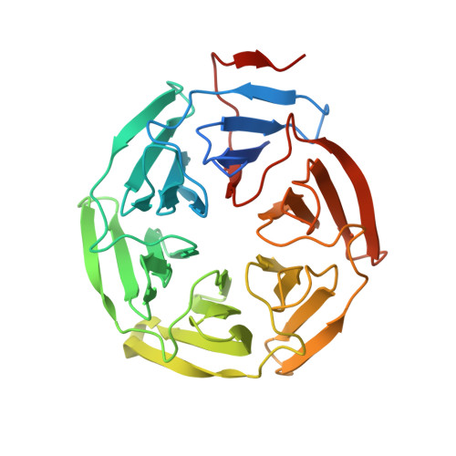
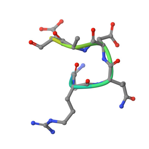
Different electrostatic potentials define ETGE and DLG motifs as hinge and latch in oxidative stress response

Tong, K.I.Padmanabhan, B.Kobayashi, A.Shang, C.Hirotsu, Y.Yokoyama, S.Yamamoto, M.

(2007) Mol Cell Biol 27: 7511-7521

  • DOI: https://doi.org/10.1128/MCB.00753-07
  • Primary Citation of Related Structures:  
    2DYH

  • PubMed Abstract: 

    Nrf2 is the regulator of the oxidative/electrophilic stress response. Its turnover is maintained by Keap1-mediated proteasomal degradation via a two-site substrate recognition mechanism in which two Nrf2-Keap1 binding sites form a hinge and latch. The E3 ligase adaptor Keap1 recognizes Nrf2 through its conserved ETGE and DLG motifs. In this study, we examined how the ETGE and DLG motifs bind to Keap1 in a very similar fashion but with different binding affinities by comparing the crystal complex of a Keap1-DC domain-DLG peptide with that of a Keap1-DC domain-ETGE peptide. We found that these two motifs interact with the same basic surface of either Keap1-DC domain of the Keap1 homodimer. The DLG motif works to correctly position the lysines within the Nrf2 Neh2 domain for efficient ubiquitination. Together with the results from calorimetric and functional studies, we conclude that different electrostatic potentials primarily define the ETGE and DLG motifs as a hinge and latch that senses the oxidative/electrophilic stress.


  • Organizational Affiliation

    Graduate School of Comprehensive Human Sciences, Center for TARA, JST-ERATO Environmental Response Project, University of Tsukuba, 1-1-1 Tennoudai, Tsukuba 305-8577, Japan.


Macromolecules
Find similar proteins by:  (by identity cutoff)  |  3D Structure
Entity ID: 1
MoleculeChains Sequence LengthOrganismDetailsImage
Kelch-like ECH-associated protein 1318Mus musculusMutation(s): 0 
UniProt
Find proteins for Q9Z2X8 (Mus musculus)
Explore Q9Z2X8 
Go to UniProtKB:  Q9Z2X8
Entity Groups  
Sequence Clusters30% Identity50% Identity70% Identity90% Identity95% Identity100% Identity
UniProt GroupQ9Z2X8
Sequence Annotations
Expand
  • Reference Sequence

Find similar proteins by:  Sequence   |   3D Structure  

Entity ID: 2
MoleculeChains Sequence LengthOrganismDetailsImage
Nrf2/Neh2 peptide from Nuclear factor erythroid 2-related factor 215N/AMutation(s): 0 
UniProt & NIH Common Fund Data Resources
Find proteins for Q60795 (Mus musculus)
Explore Q60795 
Go to UniProtKB:  Q60795
IMPC:  MGI:108420
Entity Groups  
Sequence Clusters30% Identity50% Identity70% Identity90% Identity95% Identity100% Identity
UniProt GroupQ60795
Sequence Annotations
Expand
  • Reference Sequence
Experimental Data & Validation

Experimental Data

  • Method: X-RAY DIFFRACTION
  • Resolution: 1.90 Å
  • R-Value Free: 0.211 
  • R-Value Work: 0.172 
  • R-Value Observed: 0.174 
  • Space Group: P 61
Unit Cell:
Length ( Å )Angle ( ˚ )
a = 103.131α = 90
b = 103.131β = 90
c = 56.141γ = 120
Software Package:
Software NamePurpose
REFMACrefinement
DNAdata collection
HKL-2000data reduction
SCALEPACKdata scaling
MOLREPphasing

Structure Validation

View Full Validation Report


Entry History 

Deposition Data

Revision History  (Full details and data files)

  • Version 1.0: 2007-09-04
    Type: Initial release
  • Version 1.1: 2011-07-13
    Changes: Version format compliance
  • Version 1.2: 2023-10-25
    Changes: Data collection, Database references, Derived calculations, Refinement description