


Crystal structure of the putative transcriptional regulator SCO0337 from Streptomyces coelicolor A3(2)
Hayashi, T., Tanaka, Y., Sakai, N., Yao, M., Tamura, T., Tanaka, I.To be published.
Experimental Data Snapshot
wwPDB Validation 3D Report Full Report

Entity ID: 1 | |||||
|---|---|---|---|---|---|
| Molecule | Chains | Sequence Length | Organism | Details | Image |
| putative transcriptional regulator | 195 | Streptomyces coelicolor A3(2) | Mutation(s): 2 | 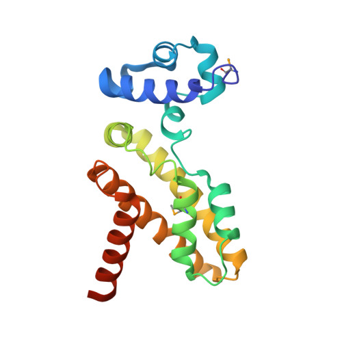 | |
UniProt | |||||
Find proteins for Q9RK42 (Streptomyces coelicolor (strain ATCC BAA-471 / A3(2) / M145)) Explore Q9RK42 Go to UniProtKB: Q9RK42 | |||||
Entity Groups | |||||
| Sequence Clusters | 30% Identity50% Identity70% Identity90% Identity95% Identity100% Identity | ||||
| UniProt Group | Q9RK42 | ||||
Sequence AnnotationsExpand | |||||
| |||||
| Modified Residues 1 Unique | |||||
|---|---|---|---|---|---|
| ID | Chains | Type | Formula | 2D Diagram | Parent |
| MSE Query on MSE | A | L-PEPTIDE LINKING | C5 H11 N O2 Se |  | MET |
| Length ( Å ) | Angle ( ˚ ) |
|---|---|
| a = 102.952 | α = 90 |
| b = 42.864 | β = 90 |
| c = 43.933 | γ = 90 |
| Software Name | Purpose |
|---|---|
| CNS | refinement |
| HKL-2000 | data reduction |
| SCALEPACK | data scaling |
| SOLVE | phasing |
RCSB PDB (citation) is hosted by
RCSB PDB is a member of the

