2BJJ

Structure of recombinant human lactoferrin produced in the milk of transgenic cows


Experimental Data Snapshot

  • Method: X-RAY DIFFRACTION
  • Resolution: 2.40 Å
  • R-Value Free: 0.231 
  • R-Value Work: 0.181 
  • R-Value Observed: 0.184 

wwPDB Validation   3D Report Full Report


Ligand Structure Quality Assessment 


This is version 1.5 of the entry. See complete history


Literature

The Protein Structure of Recombinant Human Lactoferrin Produced in the Milk of Transgenic Cows Closely Matches the Structure of Human Milk-Derived Lactoferrin

Thomassen, E.A.J.Van Veen, H.A.Van Berkel, P.H.C.Nuijens, J.H.Abrahams, J.P.

(2005) Transgenic Res 14: 397

  • DOI: https://doi.org/10.1007/s11248-005-3233-0
  • Primary Citation of Related Structures:  
    2BJJ

  • PubMed Abstract: 

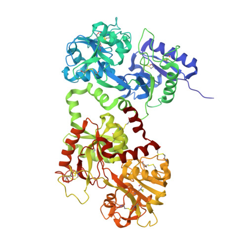
    Human lactoferrin (hLF) is an iron-binding glycoprotein involved in the host defence against infection and excessive inflammation. As the availability of (human milk-derived) natural hLF is limited, alternative means of production of this biopharmaceutical are extensively researched. Here we report the crystal structure of recombinant hLF (rhLF) expressed in the milk of transgenic cows at a resolution of 2.4 A. To our knowledge, the first reported structure of a recombinant protein produced in milk of transgenic livestock. Even though rhLF contains oligomannose- and hybrid-type N-linked glycans next to complex-type glycans, which are the only glycans found on natural hLF, the structures are identical within the experimental error (r.m.s. deviation of only 0.28 A for the main-chain atoms). Of the differences in polymorphic amino acids between the natural and rhLF variant used, only the side-chain of Asp561 could be modeled into the rhLF electron density map. Taken together, the results confirm the structural integrity of the rhLF variant used in this study. It also confirms the validity of the transgenic cow mammary gland as a vehicle to produce recombinant human proteins.


  • Organizational Affiliation

    Biophysical Structural Chemistry, Leiden Institute of Chemistry, Leiden University, Einsteinweg 55, 2333 CC Leiden, The Netherlands.


Macromolecules
Find similar proteins by:  (by identity cutoff)  |  3D Structure
Entity ID: 1
MoleculeChains Sequence LengthOrganismDetailsImage
LACTOTRANSFERRINA [auth X]692Homo sapiensMutation(s): 0 
UniProt & NIH Common Fund Data Resources
Find proteins for P02788 (Homo sapiens)
Explore P02788 
Go to UniProtKB:  P02788
PHAROS:  P02788
GTEx:  ENSG00000012223 
Entity Groups  
Sequence Clusters30% Identity50% Identity70% Identity90% Identity95% Identity100% Identity
UniProt GroupP02788
Sequence Annotations
Expand
  • Reference Sequence
Experimental Data & Validation

Experimental Data

  • Method: X-RAY DIFFRACTION
  • Resolution: 2.40 Å
  • R-Value Free: 0.231 
  • R-Value Work: 0.181 
  • R-Value Observed: 0.184 
  • Space Group: P 21 21 21
Unit Cell:
Length ( Å )Angle ( ˚ )
a = 55.941α = 90
b = 97.379β = 90
c = 156.297γ = 90
Software Package:
Software NamePurpose
REFMACrefinement
MOSFLMdata reduction
SCALAdata scaling
MOLREPphasing

Structure Validation

View Full Validation Report


Ligand Structure Quality Assessment 


Entry History 

Deposition Data

Revision History  (Full details and data files)

  • Version 1.0: 2005-08-01
    Type: Initial release
  • Version 1.1: 2011-05-08
    Changes: Version format compliance
  • Version 1.2: 2011-07-13
    Changes: Version format compliance
  • Version 1.3: 2017-06-28
    Changes: Data collection
  • Version 1.4: 2020-07-29
    Type: Remediation
    Reason: Carbohydrate remediation
    Changes: Advisory, Data collection, Derived calculations, Other, Structure summary
  • Version 1.5: 2023-12-13
    Changes: Data collection, Database references, Refinement description, Structure summary