2B5V

Crystal structure of glucose dehydrogenase from Haloferax mediterranei


Experimental Data Snapshot

  • Method: X-RAY DIFFRACTION
  • Resolution: 2.00 Å
  • R-Value Free: 0.207 
  • R-Value Work: 0.161 

wwPDB Validation   3D Report Full Report


Ligand Structure Quality Assessment 


This is version 1.3 of the entry. See complete history


Literature

Analysis of protein solvent interactions in glucose dehydrogenase from the extreme halophile Haloferax mediterranei.

Britton, K.L.Baker, P.J.Fisher, M.Ruzheinikov, S.Gilmour, D.J.Bonete, M.-J.Ferrer, J.Pire, C.Esclapez, J.Rice, D.W.

(2006) Proc Natl Acad Sci U S A 103: 4846-4851

  • DOI: https://doi.org/10.1073/pnas.0508854103
  • Primary Citation of Related Structures:  
    2B5V, 2B5W

  • PubMed Abstract: 

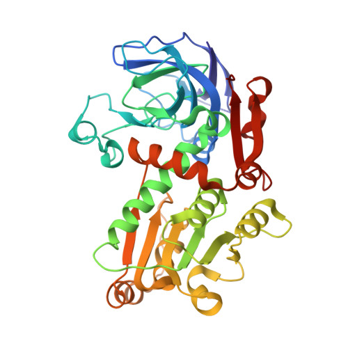
    The structure of glucose dehydrogenase from the extreme halophile Haloferax mediterranei has been solved at 1.6-A resolution under crystallization conditions which closely mimic the "in vivo" intracellular environment. The decoration of the enzyme's surface with acidic residues is only partially neutralized by bound potassium counterions, which also appear to play a role in substrate binding. The surface shows the expected reduction in hydrophobic character, surprisingly not from changes associated with the loss of exposed hydrophobic residues but rather arising from a loss of lysines consistent with the genome wide-reduction of this residue in extreme halophiles. The structure reveals a highly ordered, multilayered solvation shell that can be seen to be organized into one dominant network covering much of the exposed surface accessible area to an extent not seen in almost any other protein structure solved. This finding is consistent with the requirement of the enzyme to form a protective shell in a dehydrating environment.


  • Organizational Affiliation

    Krebs Institute for Biomolecular Research, Department of Molecular Biology and Biotechnology, University of Sheffield, Sheffield S10 2TN, United Kingdom.


Macromolecules
Find similar proteins by:  (by identity cutoff)  |  3D Structure
Entity ID: 1
MoleculeChains Sequence LengthOrganismDetailsImage
glucose dehydrogenase357Haloferax mediterraneiMutation(s): 0 
Gene Names: gdh
EC: 1.1.1.47
UniProt
Find proteins for Q977U7 (Haloferax mediterranei (strain ATCC 33500 / DSM 1411 / JCM 8866 / NBRC 14739 / NCIMB 2177 / R-4))
Explore Q977U7 
Go to UniProtKB:  Q977U7
Entity Groups  
Sequence Clusters30% Identity50% Identity70% Identity90% Identity95% Identity100% Identity
UniProt GroupQ977U7
Sequence Annotations
Expand
  • Reference Sequence
Small Molecules
Ligands 1 Unique
IDChains Name / Formula / InChI Key2D Diagram3D Interactions
NAP
Query on NAP

Download Ideal Coordinates CCD File 
B [auth A]NADP NICOTINAMIDE-ADENINE-DINUCLEOTIDE PHOSPHATE
C21 H28 N7 O17 P3
XJLXINKUBYWONI-NNYOXOHSSA-N
Experimental Data & Validation

Experimental Data

  • Method: X-RAY DIFFRACTION
  • Resolution: 2.00 Å
  • R-Value Free: 0.207 
  • R-Value Work: 0.161 
  • Space Group: I 2 2 2
Unit Cell:
Length ( Å )Angle ( ˚ )
a = 61.791α = 90
b = 110.225β = 90
c = 152.332γ = 90
Software Package:
Software NamePurpose
DENZOdata reduction
SCALEPACKdata scaling
MLPHAREphasing
REFMACrefinement
CNSrefinement

Structure Validation

View Full Validation Report


Ligand Structure Quality Assessment 


Entry History 

Deposition Data

Revision History  (Full details and data files)

  • Version 1.0: 2006-04-04
    Type: Initial release
  • Version 1.1: 2008-05-01
    Changes: Version format compliance
  • Version 1.2: 2011-07-13
    Changes: Derived calculations, Version format compliance
  • Version 1.3: 2024-02-14
    Changes: Data collection, Database references, Derived calculations