2B55

Human cyclin dependent kinase 2 (cdk2) complexed with indenopyraxole DIN-101312


Experimental Data Snapshot

  • Method: X-RAY DIFFRACTION
  • Resolution: 1.85 Å
  • R-Value Free: 0.256 
  • R-Value Work: 0.213 

wwPDB Validation   3D Report Full Report


Ligand Structure Quality Assessment 


This is version 1.3 of the entry. See complete history


Literature

Synthesis and Evaluation of Indenopyrazoles as Cyclin-Dependent Kinase Inhibitors. 3. Structure Activity Relationships at C3

Yue, E.W.Higley, C.A.Dimeo, S.V.Carini, D.J.Nugiel, D.A.Benware, C.Benfield, P.A.Cox, S.Burton, C.R.Grafstrom, R.H.Sharp, D.M.Sisk, L.M.Boylan, J.F.Muckelbauer, J.K.Smallwood, A.M.Chen, H.Chang, C.-H.Seitz, S.P.Trainor, G.L.

(2002) J Med Chem 45: 5233-5248

  • DOI: https://doi.org/10.1021/jm0201722
  • Primary Citation of Related Structures:  
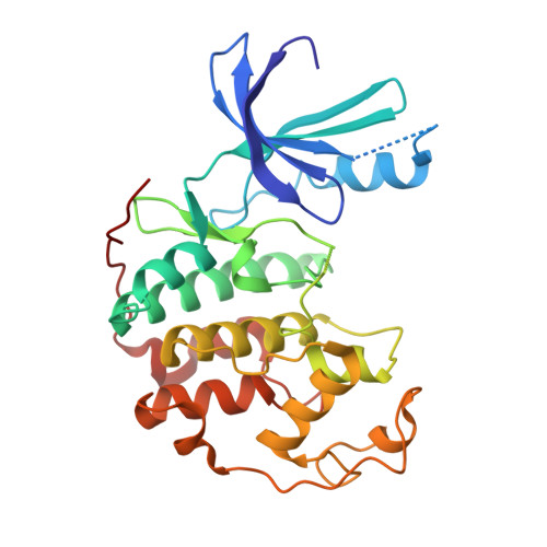
    2B55

  • PubMed Abstract: 

    The identification of indeno[1,2-c]pyrazol-4-ones as inhibitors of cyclin-dependent kinases (CDKs) has led to the discovery of a series of novel and potent compounds. Herein, we report the effects of substitutions at C3 of the indeno[1,2-c]pyrazol-4-one core with alkyls, heterocycles, and substituted phenyls. Substitutions at the para position of the phenyl ring at C3 were generally well-tolerated; however, larger groups were generally inactive. For alkyls directly attached to C3, longer chain substituents were not tolerated; however, shorter alkyl groups and cyclic alkyls were acceptable. In general, the heterocycles at C3 gave the most potent analogues. One such heterocycle, 24j, was examined in detail and was determined to have a biological profile consistent with CDK inhibition. An X-ray crystal structure of one of the alkyl compounds, 13q, complexed with CDK2 was determined and showed the inhibitor residing in the adenosine 5'-triphosphate pocket of the enzyme.


  • Organizational Affiliation

    Bristol-Myers Squibb Company, Experimental Station, P.O. Box 80500, Wilmington, Delaware 19880-0500, USA. eddy.yue@bms.com


Macromolecules
Find similar proteins by:  (by identity cutoff)  |  3D Structure
Entity ID: 1
MoleculeChains Sequence LengthOrganismDetailsImage
Cell division protein kinase 2298Homo sapiensMutation(s): 0 
EC: 2.7.1.37
UniProt & NIH Common Fund Data Resources
Find proteins for P24941 (Homo sapiens)
Explore P24941 
Go to UniProtKB:  P24941
PHAROS:  P24941
GTEx:  ENSG00000123374 
Entity Groups  
Sequence Clusters30% Identity50% Identity70% Identity90% Identity95% Identity100% Identity
UniProt GroupP24941
Sequence Annotations
Expand
  • Reference Sequence
Small Molecules
Ligands 1 Unique
IDChains Name / Formula / InChI Key2D Diagram3D Interactions
D31
Query on D31

Download Ideal Coordinates CCD File 
B [auth A]2-(4-(AMINOMETHYL)PIPERIDIN-1-YL)-N-(3_CYCLOHEXYL-4-OXO-2,4-DIHYDROINDENO[1,2-C]PYRAZOL-5-YL)ACETAMIDE
C24 H31 N5 O2
AITZHKQVQNLKHI-UHFFFAOYSA-N
Binding Affinity Annotations 
IDSourceBinding Affinity
D31 PDBBind:  2B55 IC50: 96 (nM) from 1 assay(s)
BindingDB:  2B55 IC50: 96 (nM) from 1 assay(s)
Binding MOAD:  2B55 IC50: 96 (nM) from 1 assay(s)
Experimental Data & Validation

Experimental Data

  • Method: X-RAY DIFFRACTION
  • Resolution: 1.85 Å
  • R-Value Free: 0.256 
  • R-Value Work: 0.213 
  • Space Group: P 21 21 21
Unit Cell:
Length ( Å )Angle ( ˚ )
a = 71.98α = 90
b = 72.2β = 90
c = 53.52γ = 90
Software Package:
Software NamePurpose
DENZOdata reduction
SCALEPACKdata scaling
EPMRphasing
CNXrefinement

Structure Validation

View Full Validation Report


Ligand Structure Quality Assessment 


Entry History 

Deposition Data

Revision History  (Full details and data files)

  • Version 1.0: 2005-10-11
    Type: Initial release
  • Version 1.1: 2008-05-01
    Changes: Version format compliance
  • Version 1.2: 2011-07-13
    Changes: Version format compliance
  • Version 1.3: 2024-02-14
    Changes: Data collection, Database references, Derived calculations